Examen neurologique des membres supérieurs
Révision par les pairs par le Dr Colin Tidy, MRCGPDernière mise à jour par le Dr Hayley Willacy, FRCGP Dernière mise à jour le 1er décembre 2021
Répond aux besoins du patient lignes directrices éditoriales
- TéléchargerTélécharger
- Partager
- Langue
- Discussion
Professionnels de la santé
Les articles de référence professionnelle sont destinés aux professionnels de la santé. Ils sont rédigés par des médecins britanniques et s'appuient sur les résultats de la recherche ainsi que sur les lignes directrices britanniques et européennes. Vous trouverez peut-être l'un de nos articles sur la santé plus utile.
Dans cet article :
Il existe plus d'une façon de procéder à un examen neurologique et le clinicien doit développer sa propre technique. Une mauvaise technique ne permettra pas d'obtenir des signes ou produira des résultats erronés.
See also the separate Neurological History and Examination article which covers the basic principles of examination and technique.
Examination of the upper limbs may be performed more easily with the patient sitting in a chair or standing.
Inspection of the upper limbs1
Note if there appears to be any damage to the hands.
On inspection, note the following:
The resting posture. Note whether there is unusual rotation or clawing of the hand and whether the patient is symmetrical.
Look for muscle wasting or hypertrophy. Note whether it is focal or diffuse.
Recherchez des mouvements involontaires tels que des tremblements, des tics, des secousses myocloniques, de la chorée ou de l'athétose.
Look for muscle fasciculation (sign of lower motor neurone disease process). These are subcutaneous twitches over a muscle belly at rest. Tapping the belly may stimulate fasciculation.
Poursuivre la lecture ci-dessous
Upper limb examination of the sensory system
Examination of each of the sensory modalities1 :
Toucher léger
Utilisez le toucher léger d'un doigt, d'un morceau de coton ou d'un morceau de papier de soie.
Il est important de toucher et non de caresser, car une sensation de mouvement, comme le frottement et le grattage, est conduite le long des voies de la douleur.
Demandez au patient de fermer les yeux et de vous dire quand il sent que vous le touchez.
Comparez chaque membre dans la même position.
Le moment de chaque toucher doit être irrégulier afin d'éviter toute anticipation de la part du patient.
A logical progression is required. You may want to start testing over the shoulder and to move along the lateral aspect of the arm and up the medial side, as this moves progressively from C4 to T3 dermatomes.
Notez toute zone d'hypoesthésie ou de dysesthésie.
Toucher vif (piqûre d'épingle)
Testez en utilisant une aiguille jetable dédiée. Une aiguille hypodermique jetable est trop pointue.
Utilisez la zone sternale pour établir une base de référence pour la netteté avant de commencer.
Follow the same progression as with light touch with the patient's eyes closed, comparing both upper limbs.
Demandez au patient d'indiquer s'il s'agit d'une hypoesthésie (sensation d'émoussement) ou d'une hyperesthésie (sensation d'acuité).
Température
Ce point est souvent négligé, mais il peut être important.
An easy and practical approach is to touch the patient with a tuning fork as the metal feels cold.
Comparez la qualité de la sensation de température sur les bras, le visage, le tronc, les mains, les jambes et les pieds.
Des récipients contenant de l'eau chaude et de l'eau froide peuvent être utilisés pour une évaluation plus précise. Demandez au patient de faire la distinction entre le chaud et le froid sur différentes zones de la peau, les yeux fermés.
Sensation de la position des articulations (proprioception)
Test at the distal interphalangeal joint of the index finger.
Hold the middle phalanx with one thumb and finger and hold the medial and lateral sides of the distal phalanx with the other. Move the distal phalanx up and down, showing the patient the movement first.
Demandez au patient de fermer les yeux et de bouger la phalange distale de haut en bas de façon aléatoire. Demandez au patient de vous indiquer la direction du mouvement à chaque fois.
Test on both hands.
If there is an abnormality, move backwards to the proximal interphalangeal joint and so on until joint position sense is normal.
Sensation de vibration
Utilisez un diapason de 128 Hz et assurez-vous que le diapason vibre.
Placez-le d'abord sur le sternum pour que le patient puisse ressentir la sensation.
Then place it on one of the distal interphalangeal joints of one of the fingers.
If no vibration is sensed, move backwards to the metacarpophalangeal joint, the wrist, etc.
Demander au patient de vous dire quand le diapason cesse de vibrer peut être utile en cas de doute sur l'intégrité de son sens vibratoire.
Two-point discrimination
There are specific two-point discriminators available. If you don't have one, use a paper clip that you can open out.
Ask the patient to close their eyes.
Take the patient's index finger in one of your hands.
Using the discriminator or paper clip, touch the pulp of the finger with either one or two of the testing tips.
The patient must tell you whether they can feel one or two stimuli.
Find the minimum distance at which they can discriminate the two tips. Normal is at 3-5 mm.
Compare both index fingers and repeat for both thumbs.
Upper limb examination of the motor system2
Tonalité
This is the resistance felt when a joint is moved passively through its normal range of movement:
Ask the patient to let their shoulders and arms 'go floppy'.
Flex and extend their shoulder passively and feel for abnormality of tone.
Repeat for the elbow and wrist.
Hypertonia is found in upper motor neurone lesions; hypotonia is found in lower motor neurone lesions and cerebellar disorders.
Cogwheel rigidity may be found in Parkinson's disease.
Puissance
Une évaluation solide de la puissance est nécessaire.
Le Medical Research Council (MRC) a mis en place un système de classification recommandé pour la puissance (voir tableau). Ce système s'est avéré fiable, bien que des doutes aient été exprimés quant à l'étendue de la gamme de grades 43 .
Le test musculaire manuel est noté différemment avec 4 - Bon : ROM complet contre la gravité avec une résistance modérée, et 5 - Normal : ROM complet contre la gravité avec une résistance maximale.4 .
Ask the patient to contract the muscle group being tested and then you as the examiner try to overpower that group.
Testez les éléments suivants :
Abduction, adduction, flexion and extension of the shoulder.
Flexion and extension of the elbow.
Flexion and extension of the wrist.
Supination and pronation of the forearm.
Extension of the fingers at the metacarpophalangeal and interphalangeal joints.
Flexion, extension, adduction and abduction of the fingers and thumbs.
Échelle MRC pour la puissance musculaire
0 | Aucune contraction musculaire n'est visible. |
1 | La contraction musculaire est visible mais il n'y a pas de mouvement de l'articulation. |
2 | Les mouvements actifs des articulations sont possibles grâce à l'élimination de la pesanteur. |
3 | Le mouvement peut vaincre la gravité, mais pas la résistance de l'examinateur. |
4 | Le groupe musculaire peut vaincre la gravité et se déplacer contre une certaine résistance de la part de l'examinateur. |
5 | Puissance totale et normale contre résistance. |
Réflexes tendineux profonds
Veillez à ce que le patient soit à l'aise et détendu et que vous puissiez voir le muscle testé.
Utilisez un marteau à tendon pour frapper le tendon du muscle et observez la contraction musculaire.
Comparez les deux côtés.
Les réflexes peuvent être hyperactifs (+++), normaux (++), lents (+) ou absents (-). ± est utilisé lorsque le réflexe n'est présent qu'en cas de renforcement (voir ci-dessous).
In the upper limbs:
Test the biceps jerk (C5, C6): with their arm relaxed, hold the patient's elbow between your thumb and remaining fingers, your thumb being anterior and directly over the biceps tendon. Ideally the elbow should be held at 90°. Elicit the reflex by tapping on your thumb.
Test the triceps jerk (C6, C7): with their arm relaxed, hold the patient's arm across their lower chest/upper abdomen with one of your hands. Elicit the reflex by tapping over the triceps tendon just above and behind their elbow.
Test the supinator jerk (C5, C6): ask the patient to relax their arm across their abdomen. Elicit the reflex by tapping over the supinator tendon just above the wrist.
Test the finger jerk: with their hand relaxed, place the tips of your index and middle fingers across the palmar surface of the patient's proximal phalanges. Tap your fingers lightly with the tendon hammer. There should be slight flexion of the patient's fingers. If there is hyperreflexia, this flexion is exaggerated.
Test the Hoffmann's reflex: rest the distal interphalangeal joint of the patient's middle finger on the side of your right index finger. Use the tip of your right thumb to flick down on the patient's middle fingertip. Watch for any movement of the patient's thumb as their fingertip springs back up. Normally there is no movement; in hyperreflexia, thumb flexion can be seen.
If a reflex is difficult to elicit, try 'reinforcement' (the Jendrassik manoeuvre). Ask the patient to clench their teeth or squeeze their knees together while you try to elicit the reflexes again.
Interprétation
Les lésions du motoneurone supérieur produisent généralement une hyperréflexie.
Les lésions du motoneurone inférieur entraînent généralement une diminution ou une absence de réponse.
Isolated loss of a reflex can point to a radiculopathy affecting that segment - eg, loss of biceps jerk if there is a C5-C6 disc prolapse.
Examen de la coordination
The cerebellum helps in the co-ordination of voluntary, automatic and reflex movement. Tests of cerebellar function, however, are only valid if power and tone are normal, and that failure to perform them may also be related to power and tone abnormalities in the upper limb rather than a cerebellar problem. These include:
The finger-nose test:
The patient should keep their eyes open.
Hold one of your fingertips up in front of, and a short distance (about 30-40 cm) from, the patient.
Ask the patient to touch the tip of their nose and then to touch your fingertip alternately and repeatedly. You can continuously change your fingertip position to make the test more difficult.
You can then test for sensory ataxia by asking the patient to close their eyes and to touch the tip of their nose using their outstretched finger.
Repeat these tests on the other side.
Look for intention tremor and past-pointing as the patient touches the examiner's fingertip, which can indicate disease of the cerebellar hemispheres.
Rapid alternating movement:
The patient needs to have one palm facing upwards.
They need to touch this palm with the palmar and then dorsal sides of the fingertips of the other hand as quickly as possible. Note that they must lift the second hand between each movement and touch the same point on the other palm without rolling the hand.
Test both sides. It is normal for the dominant hand to be a little faster at this test.
Look for dysdiadochokinesis. This is inco-ordination or slow movement when trying to perform this test.
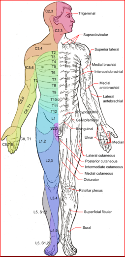
Examen neurologique

Häggström, Mikael (2014). "Galerie médicale de Mikael Häggström 2014". WikiJournal de la médecine 1 (2). DOI:10.15347/wjm/2014.008. ISSN 2002-4436. Domaine public
A note about sensation in the hand
The hand may require more intensive testing. It may be useful to return to it after testing the rest of the arm.
Test sensation on both the palmar and the dorsal aspects.
Be aware of the distribution of the median, ulnar and radial nerves:
The radial nerve supplies sensation to the skin on most of the dorsum of the hand.
The ulnar nerve supplies sensation to the palmar aspect of the little finger and the palmar aspect of the medial half of the ring finger. It also supplies the distal half of the dorsal aspect of these fingers.
The median nerve supplies sensation to the palmar aspect of the thumb, index and middle fingers and the lateral half of the ring finger. It also supplies the distal half of the dorsal aspect of these fingers.
Interprétation des résultats
The site of any lesion can be determined by looking at the pattern of any dysfunction found. The dermatomal (segmental) and peripheral nerve innervation is labelled in the diagram above.
All of the sensory modalities can be affected in peripheral neuropathies and nerve injuries, cervical radiculopathy and spinal injuries.
Si un nerf ou une racine sensorielle est touché, toutes les modalités sensorielles peuvent être réduites.
If there is a spinal cord lesion, there may not be equal diminution across all of the sensory modalities: light touch, vibration and joint position sense may remain intact while sharp touch and temperature are lost. This is because the lateral spinothalamic pathways may be damaged while the dorsal columns remain intact. Cervical syringomyelia is an example where this may happen.
Problems with joint position sense or vibration usually occur distally first.
En cas de neuropathie périphérique ou de myélopathie affectant les colonnes dorsales, la perception des vibrations peut être perdue avant la perception de la position de l'articulation.
Parietal lobe lesions can also cause impairment of two-point discrimination.
Les parties distales des membres ont tendance à être affectées dans la polyneuropathie, les jambes étant généralement touchées avant les bras. L'effet "gants et bas" se produit.
Autres lectures et références
- Examen neurologiqueExamen médical d'Oxford (OME)
- Shahrokhi M, Asuncion RMDExamen neurologique
- Compston AAides à l'investigation des lésions nerveuses périphériques. Conseil de la recherche médicale : Comité de recherche sur les lésions nerveuses. His Majesty's Stationery Office : 1942 ; pp. 48 (iii) et 74 figures et 7 diagrammes ; avec des aides à l'examen du système nerveux périphérique. Par Michael O'Brien pour les garants de Brain. Saunders Elsevier : 2010 ; pp. [8] 64 et 94 figures. Brain. 2010 Oct;133(10):2838-44. doi : 10.1093/brain/awq270.
- Naqvi U, Sherman AlClassement de la force musculaire
Poursuivre la lecture ci-dessous
Historique de l'article
Les informations contenues dans cette page sont rédigées et évaluées par des cliniciens qualifiés.
Prochaine révision prévue : 30 novembre 2026
1 Décembre 2021 | Dernière version

Demandez, partagez, connectez-vous.
Parcourez les discussions, posez des questions et partagez vos expériences sur des centaines de sujets liés à la santé.

Vous ne vous sentez pas bien ?
Évaluez gratuitement vos symptômes en ligne