Dermatofibrome
Révision par les pairs par le Dr Toni Hazell, MRCGPDernière mise à jour par Dr Rosalyn Adleman, MRCGPDernière mise à jour : 14 mars 2025
Répond aux besoins du patient lignes directrices éditoriales
- TéléchargerTélécharger
- Partager
- Langue
- Discussion
Professionnels de la santé
Les articles de référence professionnelle sont destinés aux professionnels de la santé. Ils sont rédigés par des médecins britanniques et s'appuient sur les résultats de la recherche ainsi que sur les lignes directrices britanniques et européennes. Vous trouverez peut-être l'un de nos articles sur la santé plus utile.
Dans cet article :
Synonyme : Histiocytome fibreux
Poursuivre la lecture ci-dessous
Qu'est-ce qu'un dermatofibrome ?
Les dermatofibromes sont des tumeurs cutanées fréquentes et bénignes, mais qui suscitent souvent l'inquiétude lors de leur découverte.
Causes des dermatofibromes (étiologie)
Traditionnellement, les dermatofibromes étaient attribués à une réaction à un traumatisme tel qu'une piqûre d'insecte. Cependant, l'étiologie précise n'est pas claire. Certains pensent qu'il s'agit de néoplasmes bénins plutôt que d'une réaction.
Les dermatofibromes les plus courants contiennent un mélange de fibroblastes, de macrophages et de vaisseaux sanguins. La plupart d'entre eux touchent le derme et peuvent s'étendre au sous-cutané. Un certain nombre de variantes moins courantes où l'histologie diffère, telles que l'histiocytome fibreux anévrismal, l'histiocytome fibreux hémosidérotique, l'histiocytome fibreux cellulaire et l'histiocytome fibreux épithélioïde, ont été décrites.1 Les différents types sont associés à des comportements et des résultats différents.
Poursuivre la lecture ci-dessous
Quelle est la fréquence des dermatofibromes ? (Epidémiologie) 23
Ils sont fréquents, mais il est difficile d'en déterminer l'incidence globale car la plupart des patients sont asymptomatiques et ne consultent pas de médecin.
Elles sont plus fréquentes chez les femmes que chez les hommes.
Elles peuvent survenir à tout âge, le plus souvent chez le jeune adulte.
Elles sont présentes dans toutes les ethnies.
Elles sont plus fréquentes et plus nombreuses chez les personnes souffrant d'immunosuppression.
Symptômes des dermatofibromes (présentation)3 4
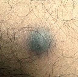
Les dermatofibromes sont généralement des nodules uniques qui se développent sur une extrémité, le plus souvent la partie inférieure des jambes. Il s'agit de nodules fermes ou durs de 0,5 à 1 cm de diamètre, qui se déplacent librement.
La surface de la peau est généralement lisse, parfois écailleuse. La peau sus-jacente peut être attachée, ce qui provoque des fossettes lorsqu'elle est pincée. La couleur de la peau varie de la couleur de la peau au rose/rouge, à la crème/blanc et au brun.
Les dermatofibromes peuvent apparaître sur n'importe quel site de la peau et les individus peuvent présenter plusieurs lésions (jusqu'à 15).
Des variantes multiples ont tendance à apparaître lorsque l'immunité est affaiblie (par exemple, maladie auto-immune, lupus érythémateux disséminé, VIH, leucémie).
Le nodule est généralement asymptomatique, mais il peut provoquer des démangeaisons ou être sensible.
Après la croissance initiale, leur taille tend à rester statique.
Dermatofibrome - vue rapprochée

© Mohammad2018, CC BY-SA 4.0, via Wikimedia Commons
Par Mohammad2018, CC BY-SA 4.0via Wikimedia Commons
Poursuivre la lecture ci-dessous
Diagnostic des dermatofibromes
Le diagnostic est généralement simple à condition de palper la lésion, car peu d'autres lésions cutanées sont aussi fermes.
Le test du pincement est utile (mais pas définitif) : en pressant la lésion sur les côtés, on obtient un capitonnage de la peau sus-jacente.
Au dermatoscope, les dermatofibromes présentent généralement un réseau pigmentaire et une tache blanche centrale, mais les variations sont considérables.5 Une étude a noté une différence d'aspect à la dermatoscopie en fonction de la localisation de la lésion.6
La biopsie d'excision est utile lorsque l'incertitude diagnostique persiste après l'examen.
Diagnostic différentiel
Comprend :
Cicatrice chéloïde ou hypertrophique.
Carcinome métastatique de la peau.
Naevus de Spitz.
Les dermatofibromes pénétrants profonds peuvent être difficiles à distinguer, même histologiquement, de rares tumeurs fibrohistiocytaires malignes, comme le dermatofibrosarcome protuberans.7
Prise en charge des dermatofibromes
Rassurer - en général, aucun traitement n'est nécessaire.
Retirer en cas de désagrément esthétique, de symptôme ou d'incertitude diagnostique. Il convient de noter que le taux de récidive locale est important.
L'excision elliptique ou la biopsie à l'emporte-pièce donnent généralement les résultats les plus satisfaisants : l'excision par rasage ou la cryothérapie présentent des risques plus élevés d'excision incomplète et de récidive.
Une étude néerlandaise a montré qu'environ 2 % des excisions sous-cutanées réalisées par des médecins généralistes donnaient lieu à des tumeurs malignes rares ou inattendues, et des études britanniques ont montré un manque de fiabilité dans le diagnostic des tumeurs malignes cutanées par les médecins généralistes.8 9 Par conséquent, même si l'excision est pratiquée pour des raisons esthétiques ou symptomatiques, il vaut la peine d'envoyer des échantillons à l'histologie.
Quand référer
L'orientation vers un spécialiste n'est normalement indiquée qu'en cas d'incertitude diagnostique, afin de faire la distinction avec d'autres lésions pigmentaires potentiellement nocives.
Pronostic
Les dermatofibromes sont presque toujours bénins - des cas extrêmement rares de dermatofibromes cellulaires métastasés ont été rapportés, bien que la distinction histologique avec d'autres tumeurs puisse s'avérer difficile.10 Assurer le suivi des lésions histologiquement atypiques ou récidivantes.
La plupart sont statiques et persistent indéfiniment, bien qu'il soit rare qu'elles régressent spontanément.
Le type cellulaire a tendance à grossir et, comme nous l'avons vu plus haut, on a parfois observé des métastases.
Ils peuvent être irrités de manière répétée, par exemple par le rasage.
Autres lectures et références
- Higgins JC, Maher MH, Douglas MSDiagnostic des tumeurs cutanées bénignes courantes. Am Fam Physician. 2015 Oct 1;92(7):601-7.
- Marghoob AA, Usatine RP, Jaimes NDermoscopie pour le médecin de famille. Am Fam Physician. 2013 Oct 1;88(7):441-50.
- Myers DJ, Fillman EPDermatofibrome. StatPearls, septembre 2021.
- Dermatofibrome; DermIS.
- Alves JV, Matos DM, Barreiros HF, et al.Variantes de dermatofibrome--une étude histopathologique. An Bras Dermatol. 2014 May-Jun;89(3):472-7.
- Myers DJ, Fillman EP. Dermatofibrome. [Mis à jour le 29 février 2024]. In : StatPearls [Internet]. Treasure Island (FL) : StatPearls Publishing; 2025 Jan
- DermatofibromeSociété de dermatologie en soins primaires
- DermatofibromeDermNet NZ
- Zaballos P, Puig S, Llambrich A, et alDermoscopie des dermatofibromes : étude morphologique prospective de 412 cas. Arch Dermatol. 2008 Jan;144(1):75-83.
- Brancaccio G, Nuzzo T, Di Maio R, et alLe Dermatofibrome a un aspect dermoscopique différent sur le tronc et les extrémités. G Ital Dermatol Venereol. 2015 Dec 23.
- Hanly AJ, Jorda M, Elgart GW, et alHigh proliferative activity excludes dermatofibroma : report of the utility of MIB-1 in the differential diagnosis of selected fibrohistiocytic tumors (Activité proliférative élevée excluant le dermatofibrome : rapport sur l'utilité de MIB-1 dans le diagnostic différentiel de certaines tumeurs fibrohistiocytaires). Arch Pathol Lab Med. 2006 Jun;130(6):831-4.
- Buis PA, Verweij W, van Diest PJValeur de l'analyse histopathologique des excisions sous-cutanées par les médecins généralistes. BMC Fam Pract. 2007 Jan 26;8:5.
- George S, Pockney P, Primrose J, et alA prospective randomised comparison of minor surgery in primary and secondary care. L'essai MiSTIC. Health Technol Assess. 2008 mai;12(23):iii-iv, ix-38.
- Bandarchi B, Ma L, Marginean C, et alD2-40, un nouveau marqueur immunohistochimique pour différencier le dermatofibrome du dermatofibrosarcome protuberans. Mod Pathol. 2010 Mar;23(3):434-8. Epub 2010 Jan 8.
Poursuivre la lecture ci-dessous
Historique de l'article
Les informations contenues dans cette page sont rédigées et évaluées par des cliniciens qualifiés.
Prochaine révision prévue : 13 mars 2028
14 mars 2025 | Dernière version

Demandez, partagez, connectez-vous.
Parcourez les discussions, posez des questions et partagez vos expériences sur des centaines de sujets liés à la santé.

Vous ne vous sentez pas bien ?
Évaluez gratuitement vos symptômes en ligne