Erythème noueux
Revu par des pairs par Dr Colin Tidy, MRCGPDernière mise à jour par Dr Toni Hazell, FRCGPDernière mise à jour 11 juil. 2022
Respecte les directives éditoriales
- TéléchargerTélécharger
- Partager
- Language
- Discussion
- Version audio
- Ajouter aux sources préférées sur Google
Professionnels de la santé
Les articles de référence professionnelle sont conçus pour être utilisés par les professionnels de la santé. Ils sont rédigés par des médecins britanniques et basés sur des preuves de recherche, des directives britanniques et européennes. Vous pouvez trouver le Erythème noueux article plus utile, ou l'un de nos autres articles de santé.
What is erythema nodosum?
Erythema nodosum is presumed to be a hypersensitivity reaction. It is often a dermatological manifestation of infectious, or other, disease.1
How common is erythema nodosum? (Epidemiology)
The prevalence of erythema nodosum varies from country to country. It is approximately five times more common in adult women than in adult men and the peak incidence is between the ages of 20 and 40, although it can occur at any age. In children, the incidence is equal in boys and girls.2 3
Erythema nodosum symptoms2 3
The eruptive phase of erythema nodosum begins with fever, aching and arthralgia, accompanied by a painful rash. .
Lesions begin as red, tender nodules. The borders are poorly defined and they are 2-6 cms in diameter.
In the first week of having erythema nodosum the lesions become tense, hard and painful. In the second week, they may become fluctuant, rather like an abscess but they do not suppurate or ulcerate. Individual lesions last around two weeks but occasionally, new lesions continue to appear for three to six weeks.
Aching legs and swollen ankles may persist for many weeks. In the first week they are bright red but in the second week there is a blue or purple hue, even turning yellow like a resolving bruise before disappearing in a couple of weeks.
They can occur anywhere but are usually on the anterior aspect of the lower leg.
Erythema nodosum on legs
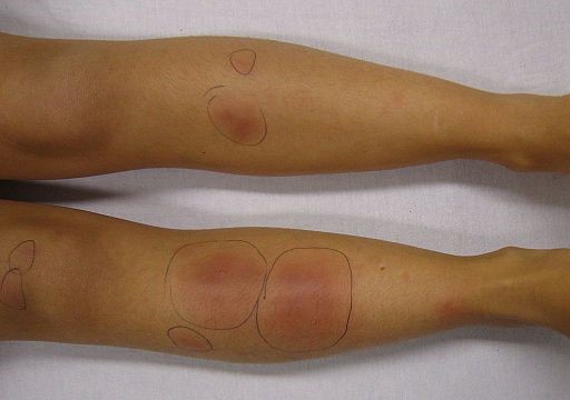
© James Heilman, MD, CC BY-SA 3.0, via Wikimedia Commons
When the aetiology is an infection the lesions usually heal in six to eight weeks but 30% of idiopathic cases last six months. Arthralgia occurs in more than half of patients and begins either during the eruptive phase or two to four weeks before. Joints are red, swollen and tender, sometimes with effusions. Morning stiffness may occur. The ankles, knees and wrists are most often involved. Synovitis resolves in a few weeks but joint pain and stiffness may last up to six months. There are no destructive changes in the joint and synovial fluid is acellular and the rheumatoid factor is negative.
Associated diseases and causes1 3 4
Erythema nodosum is often indicative of an underlying infectious disease but in up to 72% of patients a cause is never found. Some underlying causes are not infectious.
The most common causes are streptococcal infection, primary TB, Behçet's disease, drug reactions, pregnancy and inflammatory bowel disease.
Sarcoid can also cause erythema nodosum.
Leprosy can produce a clinical picture of erythema nodosum, although the histological picture of the lesions is different.
Various forms of gastroenteritis - especially Yersinia enterocolitica, Salmonella spp. and Campylobacter spp. - can be associated.
Lymphogranuloma venereum may be a cause.
Mycoplasma pneumonia can be associated.
Fungal infections are less common in the UK but coccidioidomycosis is important in Southwest USA. It may occur in histoplasmosis and blastoplasmosis.
Sulfonamides are used less often nowadays but other drugs to be implicated include sulfonylureas, gold and oral contraceptives.
It can precede the diagnosis of Hodgkin's lymphoma and non-Hodgkin's lymphoma by months.
In pregnancy it is usually seen in the second trimester. It is likely to recur in future pregnancies and may occur with oral contraceptives.
There are rare cases (<1 in 100) associated with Epstein-Barr virus, hepatitis B and hepatitis C and HIV.
Enquêtes3 4
Although in many cases it is idiopathic, it is important to exclude serious underlying disease:
A throat swab for streptococcus is an appropriate test, although it may well be negative, even with streptococcal disease.
Anti-streptococcal O (ASO) titre may be more helpful, although a normal titre does not exclude infection. A rising titre may be more valuable.
Arrange an FBC and ESR. ESR is often very high regardless of the aetiology, and CRP may be more contributory.
Stool examination for Y. enterocolitica, Salmonella spp. and Campylobacter spp. may yield results, as may blood cultures - however, most laboratories will not process formed stool for infection so this is generally only worth doing if the patient has diarrhoea.
In sarcoidosis, calcium and angiotensin-converting enzyme (ACE) are often raised.5
CXR may show bilateral hilar lymphadenopathy (BHL) in sarcoidosis, unilateral or asymmetrical adenopathy in malignancy, or evidence of pulmonary tuberculosis.
Intradermal skin tests may be required to exclude tuberculosis and coccidioidomycosis.1
Excisional biopsy may be helpful where the diagnosis is in doubt.
Diagnostic différentiel3 6
If the rash lasts for more than eight weeks then it is particularly important to consider rarer causes; this is also the case if there is ulceration or if the lesions are not located on the legs, as these are atypical features for erythema nodosum.
Erythema induratum (modular vasculitis).
Vasculitides such as polyarteritis nodosa.
Rheumatoid nodule.
Thrombophlébite superficielle (standard or superficial migratory thrombophlebitis).
Erythema nodosum treatment and management7
Most cases are self-limiting and require only symptomatic relief.
If an aetiology has been discovered then appropriate therapy is in order, depending on the underlying condition.
A degree of relief can be obtained with cool compresses and bed rest with elevation of the foot of the bed. Bed rest has been advocated for many years and is anecdotally useful but the evidence base is lacking.
Non-steroidal anti-inflammatory drugs (NSAIDs) and colchicine are useful and no other drugs are usually needed. Steroids are beneficial but should be used with caution and may be contra-indicated if infection has not been excluded.
The literature on erythema nodosum lists a host of possible treatments for persisting cases - these include potassium iodide, oral steroids, tetracyclines, macrolides and biologic drugs; however, it would seem wise to involve secondary care at this point to be sure that an underlying diagnosis isn't being missed.2 3
Pronostic
Erythema nodosum usually resolves within six weeks but it may be more protracted, especially if the underlying cause of erythema nodosum remains or when it is idiopathic. Serious complications are unusual unless part of the underlying disease. Chronic or recurrent disease is rare. Lesions heal without atrophy or scarring.4
Mises à jour exclusives pour les professionnels de la santé
Restez informé des dernières mises à jour cliniques, des perspectives professionnelles et des conseils fondés sur des preuves. La newsletter Patient Pro sélectionne des contenus essentiels pour les professionnels de santé—livrés directement dans votre boîte de réception.
En vous abonnant, vous acceptez notre Politique de confidentialité. Vous pouvez vous désabonner à tout moment. Nous ne vendons jamais vos données.
Lectures complémentaires et références
- Shimizu M, Hamaguchi Y, Matsushita T, et al; Sequentially appearing erythema nodosum, erythema multiforme and Henoch-Schonlein purpura in a patient with Mycoplasma pneumoniae infection: a case report. J Med Case Rep. 2012 Nov 23;6(1):398. doi: 10.1186/1752-1947-6-398.
- Erythema Nodosum; Société de Dermatologie de Soins Primaires
- Whig J, Mahajan V, Kashyap A, et al; Erythema nodosum: Atypical presentation of common disease. Lung India. 2010 Jul;27(3):181-2. doi: 10.4103/0970-2113.68319.
- Perez-Garza DM, Chavez-Alvarez S, Ocampo-Candiani J, et al; Erythema Nodosum: A Practical Approach and Diagnostic Algorithm. Am J Clin Dermatol. 2021 May;22(3):367-378. doi: 10.1007/s40257-021-00592-w. Epub 2021 Mar 8.
- Dermnetz; Erythema Nodosum
- Schwartz RA, Nervi SJ; Erythema nodosum: a sign of systemic disease. Am Fam Physician. 2007 Mar 1;75(5):695-700.
- Fowler A, Dargan P, Jones A; Puzzling hypercalcaemia: sarcoidosis without lung involvement. J R Soc Med. 2005 Feb;98(2):60-1.
- Yi SW, Kim EH, Kang HY, et al; Erythema nodosum: clinicopathologic correlations and their use in differential diagnosis. Yonsei Med J. 2007 Aug 31;48(4):601-8.
- Passarini B, Infusino SD; Erythema nodosum. G Ital Dermatol Venereol. 2013 Aug;148(4):413-7.
À propos de l'auteurVoir la biographie complète

Dr Toni Hazell, FRCGP
MBBS, BSc, FRCGP, DFSRH, Dip GU med, DRCOG, DCH (London, UK, 2000)
Le Dr Toni Hazell a obtenu son diplôme de l'École de médecine de l'hôpital St. Mary et a effectué son VTS à l'hôpital Northwick Park.
À propos du critiqueVoir la biographie complète

Dr Colin Tidy, MRCGP
Médecin généraliste, Auteur médical
MBBS, MRCGP, MRCP (Paediatrics), DCH
Le Dr Colin Tidy est un médecin du NHS, basé dans l'Oxfordshire.
Historique de l'article
Les informations sur cette page sont rédigées et examinées par des cliniciens qualifiés.
Article également disponible en Anglais, Allemand, Espagnol, Français, Italien, Portugais, Hindi, Hébreu, Arabe, and Suédois.
Prochaine révision prévue : 10 juil. 2027
11 juil. 2022 | Dernière version

Demandez, partagez, connectez-vous.
Parcourez les discussions, posez des questions et partagez vos expériences sur des centaines de sujets de santé.

Vous ne vous sentez pas bien ?
Évaluez vos symptômes en ligne gratuitement
Plus en dermatologie
- Naevus de Becker
- Nævus bleu
- Filariose de la cavité corporelle
- Candidose
- Psoriasis en plaques chronique
- Pemphigoïde cicatricielle
- Lupus érythémateux discoïde
- Erythema induratum
- Dracunculose
- Hémangiomes de la peau
- VIH et troubles cutanés
- Kératose pilaire
- Sclérodermie localisée
- Nocardia
- Syndrome de Parkes Weber
- Infection par le parvovirus
- Dyshidrose
- Maladie des ongles psoriasiques
- Zona
- Carcinome épidermoïde de la peau