scanner CT
Preparation and complications
Revu par Dr Colin Tidy, MRCGPDernière mise à jour par Dr Rachel Hudson, MRCGPDernière mise à jour 20 juin 2023
Respecte les directives éditoriales
- TéléchargerTélécharger
- Partager
- Language
- Discussion
- Version audio
- Ajouter aux sources préférées sur Google
Remarque: les informations ci-dessous ne sont qu'un guide général. Les dispositions et la manière dont les tests sont effectués peuvent varier d'un hôpital à l'autre. Suivez toujours les instructions données par votre médecin ou votre hôpital local.
En un coup d'œil
Un scanner CT est un test de radiographie spécialisé qui crée des images claires de l'intérieur du corps.
Il peut montrer les tissus mous comme les muscles, les organes et le cerveau, ce que les radiographies ordinaires ne peuvent pas faire.
Un scanner peut aider à détecter des anomalies ou à guider des procédures comme des biopsies.
Vous devrez peut-être retirer les objets métalliques ou arrêter de manger avant le scan, selon la zone.
Le scan lui-même est indolore et prend généralement entre 5 et 30 minutes.
Les complications sont rares, mais les femmes enceintes devraient généralement éviter les scanners CT.
Le risque lié aux radiations est très faible, et les avantages l'emportent souvent sur ce risque.
What is a CT scan?
A CT scan is a specialised X-ray test. It can give quite clear pictures of the inside of your body. In particular, it can give good pictures of soft tissues of the body which do not show on ordinary X-ray pictures.
CT stands for computerised tomography. It is sometimes called a CAT scan. CAT stands for computerised axial tomography. (Sometimes the word 'computed' is used instead of 'computerised'.)
How is a CT scan done?
The CT scanner looks like a giant thick ring. Within the wall of the scanner there is an X-ray source. Opposite the X-ray source, on the other side of the ring, are X-ray detectors.
You lie on a couch which slides into the centre of the ring until the part of the body to be scanned is within the ring.
The X-ray machine within the ring rotates around your body. As it rotates around, the X-ray machine emits thin beams of X-rays through your body, which are detected by the X-ray detectors.
CT scanner
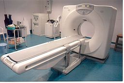
The detectors detect the strength of the X-ray beam that has passed through your body. The more dense the tissue, the less X-rays pass through. The X-ray detectors feed this information into a computer.
Different types of tissue with different densities show up as a picture on the computer monitor, in different colours or shades of grey. So, in effect, a picture is created by the computer of a slice (cross-section) of a thin section of your body.
As the couch moves slowly through the ring, the X-ray beam passes through the next section of your body. So, several cross-sectional images of the part of your body being investigated are made by the computer.
Newer scanners can even produce 3-dimensional pictures from the data received from the various slices of the part of the body being scanned.
What is a CT scan used for?
A CT scan can be done on any section of the head or body. It can give clear pictures of bones. It also gives clear pictures of soft tissues, which an ordinary X-ray test cannot show, such as muscles, organs, large blood vessels, the brain and nerves.
The most commonly performed CT scan is of the brain - to determine the cause of a stroke, or to assess serious head injuries. Other uses of a CT scan include:
To detect abnormalities in the body, such as tumours, abscesses, abnormal blood vessels, etc, when they are suspected by symptoms or other tests.
To give a surgeon a clear picture of an area of your body before certain types of surgery.
To pinpoint the exact site of tumours prior to radiothérapie.
To help doctors find the right place to take tissue samples (biopsies).
Preparing for a CT scan
Usually very little. It depends on which part of your body is to be scanned. You will be given instructions by the CT department appropriate for the scan to be done. These include:
Removing any metal objects from your body, such as jewellery, hair clips, etc.
Don't wear clothes with metal zips, studs, etc.
You may be asked not to eat or drink for a few hours before your scan.
Potentially stopping certain medicines before the procedure.
If you need an injection of contrast, as described below, it may be necessary to stop certain medicines before the procedure. This may apply to people taking metformine, a medicine used to treat diabète. If you are taking this medication your doctor should give you instructions about what to do.
Further preparations
In some situations, depending on what part of the body is being scanned, one of the following may be needed. These aim to block a certain amount of X-ray going through various tissues. This helps to give better contrast between different organs and tissues on the scan pictures.
For abdominal and pelvic scans you may be asked to have a special drink before the scan. This helps to show up the stomach and bowel more clearly.
For pelvic scans, some fluid may be put into your back passage (rectum).
For pelvic scans, women may be asked to insert a tampon into the vagina.
Sometimes a dye (contrast agent) is injected into the bloodstream via a vein in your arm. The dye may give you a flushing feeling and an odd taste in your mouth, which soon goes.
What is a CT Scan with contrast?
Sometimes you will be given a colourless substance either as a drink, as an injection, or inserted into your rectum, which will show up on the CT scan images as white (as it blocks the x-rays). This is used to show more details of structures such as blood vessels and the intestine/bowel. Contrast often contains iodine, which some people may be allergic to - if you are allergic to iodine you should inform the scan department, however you will be asked about allergies before you have the scan. If you do suffer an allergic reaction to iodine following being given it for a CT this may cause itching, wheezing or feeling sick. This is usually short lived. Rarely, a delayed allergic reaction several hours after the scan can occur which causes an itchy rash - if you have a reaction following a CT scan with contrast you should contact A&E or your GP practice.
The contrast is got rid of from your body in your urine.
Note de l'éditeur
Dr Krishna Vakharia, 1st November 2024
If you are having a scan with contrast it is also important to let the department know if you have kidney disease or have had a kidney transplant. Let them know if you are under a kidney specialist or a urologist. You may also be asked to stop certain medications such as some used for high blood pressure before a scan with contrast. Your doctor or scan department will discuss this with you if this is something you need to do.
Is a CT scan painful?
The CT scan itself is painless. You cannot see or feel X-rays. You will be asked to stay as still as possible, as otherwise the scan pictures may be blurred.
How long does a CT scan take?
Conventional CT scans can take between 5-30 minutes, depending on which part (or parts) of the body is being scanned. More modern CT scans (helical CT scans) take less than a minute and also use less radiation.
Can anybody be with me during the CT scan?
Because the scan uses X-rays, other people should not be in the same room. The operator controls the movement of the couch and scanner from behind a screen or in a separate control room. (This protects them from repeated exposure to X-rays.)
However, you can talk to them, usually via an intercom, and you will be observed at all times on a monitor. Some people feel a little anxieux or claustrophobic in the scanner room when they are on their own. A mild sedative may be offered if you are particularly anxious.
What can I expect after the CT scan?
You can return to your normal activities as soon as the scan is over. However, if you had a sedative for the scan, you will need someone to accompany you home. You will not be able to drive until the effect of the sedative wears off.
The pictures from the scan are studied by an X-ray doctor (radiologist) who sends a report to the doctor who requested the scan.
Are there any possible complications regarding a CT scan?
Complications are rare. As discussed above, some people have an allergic reaction to the dye (contrast agent) which is sometimes used. This is rare. It can be treated immediately. Very rarely, the dye may cause some kidney damage, most commonly in people already known to have kidney problems.
Femmes enceintes
If possible, pregnant women should not have a CT scan, as there is a small risk that X-rays may cause an abnormality to the unborn child.
Risks of X-ray radiation used in CT scans
CT scans use X-rays, which are a type of radiation. Exposure to large doses of radiation is linked to developing cancer or leukaemia - often many years later.
The dose of X-ray radiation needed for a CT scan is much more than for a single X-ray image but is still generally quite a low dose. The risk of harm from the dose of radiation used in CT scanning is thought to be very small but it is not totally without risk.
As a rule, the higher the dose of radiation, the greater the risk. So, for example, the larger the part of the body scanned, the greater the amount of radiation dose. And, repeat CT scans over time cause an overall increase of dose. Also, the younger you are when you have a CT scan, the greater the lifetime risk of developing cancer or leukaemia.
Various studies have aimed to estimate the risk of developing cancer or leukaemia following a CT scan (see 'Further reading' below). In general, the risk is small. In many situations, the benefit of a CT scan greatly outweighs the risk.
However, as the same study concludes: ... "although clinical benefits should outweigh the small absolute risks, radiation doses from CT scans ought to be kept as low as possible and alternative procedures, which do not involve ionising radiation, should be considered if appropriate."
Because of the small risk, the Committee on Medical Aspects of Radiation in the Environment (COMARE) has recommended that routine whole-body CT scans should not be offered to people without symptoms as part of 'health checks'. They also offer various other recommendations on the use of CT scans (see 'Further reading' below).
Sélections des patients pour Imagerie
Tests et investigations
Scintigraphie
Une scintigraphie est une méthode d'imagerie des os, des organes et d'autres parties du corps en utilisant une petite dose d'une substance radioactive. Il existe différents types de substances radioactives. Celle utilisée dépend de l'organe ou de la partie du corps à examiner. Remarque : les informations ci-dessous sont données à titre indicatif uniquement. Les modalités et la manière dont les tests sont effectués peuvent varier d'un hôpital à l'autre. Suivez toujours les instructions de votre médecin ou de l'hôpital local.
par Dr Rachel Hudson, MRCGP

Tests et investigations
Lavement baryté
Une lavement baryté est un examen utilisé pour aider à visualiser le contour du gros intestin (côlon). Remarque : les informations ci-dessous sont uniquement une ligne directrice générale. Les modalités et la manière dont les examens sont réalisés peuvent varier d’un hôpital à l’autre. Suivez toujours les instructions données par votre médecin ou votre hôpital local.
par Dr Colin Tidy, MRCGP
Questions fréquemment posées
Puis-je rentrer chez moi en voiture après avoir passé un scanner ?
Vous pouvez généralement reprendre vos activités normales immédiatement après un scanner. Cependant, si un sédatif vous a été administré pour la procédure, vous aurez besoin de quelqu'un d'autre pour vous ramener chez vous. Vous ne devez pas conduire tant que les effets du sédatif ne se sont pas complètement dissipés.
Que dois-je faire si je ressens une réaction allergique retardée au produit de contraste ?
Bien que les réactions allergiques au produit de contraste soient généralement de courte durée, une réaction retardée rare peut survenir plusieurs heures après le scan, se manifestant généralement par une éruption cutanée qui démange. Si vous ressentez cela, vous devriez contacter les urgences ou votre cabinet médical pour obtenir des conseils.
Est-il sûr de passer un scanner si j'ai des problèmes rénaux ?
Si vous souffrez d'une maladie rénale ou avez subi une greffe de rein, il est important d'informer le service de radiologie, surtout si vous passez un examen avec produit de contraste. Le produit est éliminé de votre corps par l'urine et, très rarement, il peut causer des dommages aux reins, en particulier chez ceux qui ont déjà des problèmes rénaux. Vous pourriez également avoir besoin d'arrêter temporairement certains médicaments, comme ceux pour l'hypertension, avant un examen avec contraste.
Pourquoi d'autres personnes ne peuvent-elles pas rester dans la pièce avec moi pendant un scanner CT ?
Parce que les scanners CT utilisent des rayons X, d'autres personnes ne sont pas autorisées dans la salle de scan pendant la procédure. Cela vise à les protéger d'une exposition répétée aux radiations. L'opérateur contrôlera le scanner depuis une pièce séparée ou derrière un écran de protection, mais vous serez observé sur un moniteur et pourrez communiquer avec lui via un interphone.
Existe-t-il des alternatives sans radiation aux scanners CT ?
L'article suggère que des procédures alternatives qui n'impliquent pas de rayonnement ionisant devraient être envisagées si cela est approprié. Cette recommandation est faite pour s'assurer que les doses de rayonnement des scanners CT soient maintenues aussi basses que possible, compte tenu des petits risques associés.
Lectures complémentaires et références
- Pearce MS, Salotti JA, Little MP, et al; Radiation exposure from CT scans in childhood and subsequent risk of leukaemia and brain tumours: a retrospective cohort study. Lancet. 2012 Jun 7.
- Hendee WR, O'Connor MK; Radiation risks of medical imaging: separating fact from fantasy. Radiology. 2012 Aug;264(2):312-21. doi: 10.1148/radiol.12112678.
- Comité sur les aspects médicaux des radiations dans l'environnement (COMARE)
- Meer AB, Basu PA, Baker LC, et al; Exposure to ionizing radiation and estimate of secondary cancers in the era of high-speed CT scanning: projections from the Medicare population. J Am Coll Radiol. 2012 Apr;9(4):245-50. doi: 10.1016/j.jacr.2011.12.007.
- Konda SR, Goch AM, Haglin J, et al; Ultralow-Dose CT (REDUCTION Protocol) for Extremity Fracture Evaluation Is as Safe and Effective as Conventional CT: An Evaluation of Quality Outcomes. J Orthop Trauma. 2018 May;32(5):216-222. doi: 10.1097/BOT.0000000000001137.
- Singh V, Sandean DP; Sécurité et soins des patients CT
- Cova MA, Stacul F, Quaranta R, et al; Radiological contrast media in the breastfeeding woman: a position paper of the Italian Society of Radiology (SIRM), the Italian Society of Paediatrics (SIP), the Italian Society of Neonatology (SIN) and the Task Force on Breastfeeding, Ministry of Health, Italy. Eur Radiol. 2014 Aug;24(8):2012-22. doi: 10.1007/s00330-014-3198-6. Epub 2014 May 17.
À propos de l'auteurVoir la biographie complète
Mis à jour par

Dr Rachel Hudson, MRCGP
Médecin généraliste et auteur médical
MBChB, MRCGP (2008), BSc (Medical Science), DFSRH, DRCOG, DCH
Dr Rachel Hudson est médecin généraliste du NHS travaillant dans le nord-ouest de l'Angleterre.
À propos du critiqueVoir la biographie complète

Dr Colin Tidy, MRCGP
Médecin généraliste, Auteur médical
MBBS, MRCGP, MRCP (Paediatrics), DCH
Le Dr Colin Tidy est un médecin du NHS, basé dans l'Oxfordshire.
Historique de l'article
Article également disponible en Anglais, Allemand, Espagnol, Français, Italien, Portugais, Hindi, Hébreu, Arabe, and Suédois.
Prochaine révision prévue : 12 mai 2028
20 juin 2023 | Dernière version
Partagez vos pensées ou expériences avec les autres dans notre communautés de patients.
Accès simple à des traitements fiables et à des services de santé en ligne.
Common conditions
Get help with common conditions through the NHS, without a GP appointment.





Inscrivez-vous à la newsletter Patient
Votre dose hebdomadaire de conseils de santé clairs et fiables - rédigés pour vous aider à vous sentir informé, confiant et maître de la situation.
En vous abonnant, vous acceptez notre Politique de confidentialité. Vous pouvez vous désabonner à tout moment. Nous ne vendons jamais vos données.