Scintigraphie
Isotope scan
Revu par Dr Krishna Vakharia, MRCGPDernière mise à jour par Dr Rachel Hudson, MRCGPLast updated 24 août 2023
Respecte les directives éditoriales
- TéléchargerTélécharger
- Partager
- Language
- Discussion
- Version audio
- Add to preferred sources on Google
A radionuclide scan is a way of imaging bones, organs and other parts of the body by using a small dose of a radioactive chemical. There are different types of radionuclide chemical. The one used depends on which organ or part of the body is to be scanned.
Remarque: les informations ci-dessous ne sont qu'un guide général. Les dispositions et la manière dont les tests sont effectués peuvent varier d'un hôpital à l'autre. Suivez toujours les instructions données par votre médecin ou votre hôpital local.
At a glance
A radionuclide scan uses a small amount of a radioactive chemical to create images inside your body.
The chemical is usually injected, but can be swallowed, breathed in, or given as eye drops.
Different radionuclides are used to target different organs or tissues, such as bones, kidneys, or the heart.
A special camera detects gamma rays from the chemical and converts them into pictures on a computer.
Preparation is usually minimal, but may involve stopping certain medicines or drinking fluids.
Radionuclide scans generally have no side-effects, and the radiation dose is very small.
Tell your doctor if you are pregnant, think you might be pregnant, or are breastfeeding.
Dans cet article:
Video picks for Imagerie
Continuez à lire ci-dessous
Why do you have a radionuclide scan?
A radionuclide scan may be done for all sorts of reasons. For example:
A bone scan is a common type. A radionuclide is used which collects in areas where there is a lot of bone activity (where bone cells are breaking down or repairing parts of the bone). So a bone scan is used to detect areas of bone where there is cancer, infection, or damage. These areas of activity are seen as 'hot spots' on the scan picture. See the separate leaflet called Bone Scan for more details.
A kidney scan can assess how well a kidney is working (as the radionuclide chosen is taken up by kidney cells and passes into the urine). So, the scan can detect scars on the kidney and how well urine drains from the kidney to the bladder. See the separate leaflet called DMSA Scan for more details.
Lung perfusion scan (also called a 'VQ scan') can detect blood clots in the lungs (pulmonary embolism).
A heart scan can assess blood flow to the heart muscle. Areas of poor blood flow to the heart muscle do not 'take up' the radionuclide very well and this will be shown in the picture.
A thyroid scan may be done to assess cases of overactive thyroid (hyperthyroidism). For example, some nodules (small 'lumps') are sometimes a focus of overactivity and will show as 'hot spots' on the picture. See the separate leaflet called Thyroid Scans and Uptake Tests for more details.
Lacrimal scintigraphy is done to test the function of tear ducts (lacrimal ducts.) The radionuclide is given as eye drops.
Lymphoscintigraphy is done to check the drainage of the enflés in people with a type of swelling of the legs, called lymphoedema.
There are various other types of radionuclide tests.
What preparation do I need?
Retour au sommaireThe preparation needed is usually very little. It will depend on which type of scan you are having. Your local hospital should give you specific information to help you prepare for these tests.
For some types of scan, you may be asked to have lots to drink to help to flush the radionuclide through your body.
For some types of scan you may also be asked to empty your bladder of urine before the scanning begins.
For some scans, such as thyroid scans, you may be instructed to stop certain medications for some time before the scan.
As these tests involve a small amount of radiation, pregnant women should not have them.
Remarque: let your doctor know if you are, or think you could be, pregnant. You should also let your doctor know if you are breastfeeding.
Continuez à lire ci-dessous
What happens during a radionuclide scan?
Retour au sommaireThe procedures for the different types of radionuclide scans are different. Information about your scan should be sent to you with the appointment.
Depending on the type of scan you have, you usually either swallow a small quantity of radionuclide, or it is injected into a vein in your arm.
It then takes some time - sometimes several hours (depending on what is being scanned) - for the radionuclide to travel to the target organ or tissue, and to be 'taken' into the active cells.
So, after receiving the radionuclide you may have a wait of a few hours. You may be able to go out and come back to the scanning room later in the day.
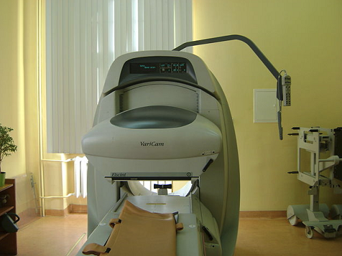
When it is time to do the scanning, you usually lie on a couch while the gamma camera detects the gamma rays coming from your body.
Gamma camera
© By Arturo 1299 (Own work) via Wikimedia Commons
The computer turns the information into a picture. You need to lie as still as possible whilst each picture is taken (so it is not blurred). Some pictures can take 20 minutes or more to expose.
The number of pictures taken and the time interval between each picture vary depending on what is being scanned. Sometimes only one picture is needed. However, for some scans (such as bone scans or heart scans), two or more pictures are needed. Each picture may be taken several hours apart. So, the whole process can take several hours.
What happens after a radionuclide scan?
Retour au sommaireRadionuclide scans do not generally cause any side-effects.
Uncommon side-effects from radionuclides may include flushing, racing heart and nausée but these are short-lived because they are flushed out of your system quickly.
Through the natural process of radioactive decay, the small amount of radioactive chemical in your body will lose its radioactivity over time. It may also pass out of your body through your urine or poo during the first few hours or days following the test.
You may be instructed to take special precautions after urinating, to flush the toilet twice and to wash your hands thoroughly. You may be advised to drink plenty of water to help flush the chemicals out of your system.
Si vous avez des contacts avec des enfants ou des femmes enceintes, vous devriez en informer votre médecin. Bien que les niveaux de radiation utilisés lors de l'examen soient faibles, ils pourraient recommander des précautions particulières. Votre hôpital devrait vous donner plus de conseils à ce sujet.
Continuez à lire ci-dessous
Are there risks with radionuclide scans?
Retour au sommaireThe term 'radioactivity' may sound alarming. But, the radioactive chemicals used in radionuclide scans are considered to be safe, and they leave the body quickly in the urine. The dose of radiation that your body receives is very small.
In many cases, the level of radiation involved is not much different to a series of a few normal X-rays. However:
Comme pour tout autre type de rayonnement (tel que la radiographie), there is a small risk that the gamma rays may affect an unborn child. So, tell your doctor if you are pregnant or if you may be pregnant.
Rarely, some people have an allergic reaction to the injected chemical. Tell your doctor if you are allergic to iodine.
Théoriquement, il est possible de faire une overdose lorsque le produit chimique est injecté. C'est très rare.
How does a radionuclide scan work?
Retour au sommaireA radionuclide (sometimes called a radioisotope or isotope) is a chemical which emits a type of radioactivity called gamma rays. A tiny amount of radionuclide is put into the body, usually by an injection into a vein. Sometimes it is breathed in, or swallowed, or given as eye drops, depending on the test.
There are different types of radionuclides. Different ones tend to collect or concentrate in different organs or tissues. So, the radionuclide used depends on which part of the body is to be scanned.
For example, if radioactive iodine is injected into a vein it is quickly taken up into the tissues of the thyroid gland. So, it is used to scan the thyroid gland.
Cells which are most 'active' in the target tissue or organ will take up more of the radionuclide. So, active parts of the tissue will emit more gamma rays than less active or inactive parts.
Gamma rays are similar to X-rays and are detected by a device called a gamma camera. The gamma rays which are emitted from inside the body are detected by the gamma camera, are converted into an electrical signal and sent to a computer.
The computer builds a picture by converting the differing intensities of radioactivity emitted into different colours or shades of grey. This is seen below in a lung perfusion scan.
Lung perfusion scan
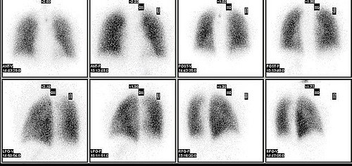
© By Myohan (Own work) via Wikimedia Commons
Alternatively areas of the target organ or tissue which emit lots of gamma rays may be shown as red spots ('hot spots') on the picture on the computer monitor.
Les zones qui émettent de faibles niveaux de rayons gamma peuvent être affichées en bleu (« zones froides »). D'autres couleurs peuvent être utilisées pour les niveaux intermédiaires d'émission de rayons gamma.
Patient picks for Imagerie

Tests et investigations
Scintigraphie au gallium
Une scintigraphie au gallium utilise une substance radioactive pour aider à créer des images pouvant montrer des zones d'infection, de blessure, d'inflammation ou de cancer possibles. Elle le fait en recherchant des zones où il y a une division cellulaire rapide dans le corps. Remarque : les informations ci-dessous sont uniquement un guide général. Les modalités et la façon dont les tests sont effectués peuvent varier d'un hôpital à l'autre. Suivez toujours les instructions données par votre médecin ou votre hôpital local.
par Dr Mary Harding, MRCGP

Tests et investigations
Scan CT
Remarque : les informations ci-dessous ne sont qu'un guide général. Les dispositions et la manière dont les tests sont effectués peuvent varier d'un hôpital à l'autre. Suivez toujours les instructions données par votre médecin ou votre hôpital local.
par Dr Rachel Hudson, MRCGP
Questions fréquemment posées
What is the purpose of a radionuclide scan and how does it help diagnose conditions?
A radionuclide scan uses a small amount of a radioactive chemical, which is introduced into your body. This chemical is designed to collect in specific organs or tissues. A special camera then detects the gamma rays emitted by this chemical. By observing how the radionuclide collects in different areas, doctors can identify problems such as inflammation, infection, tumors, or areas of poor blood flow that might not be visible with other imaging techniques.
How long will I be exposed to radiation after a radionuclide scan?
The radionuclide used in the scan has a short lifespan and loses its radioactivity over time through a natural process called radioactive decay. It also leaves your body relatively quickly, primarily through your urine or faeces, usually within the first few hours or days following the test. You might be advised to drink plenty of water to help flush it out of your system.
Can I eat or drink before my radionuclide scan?
Preparation instructions for a radionuclide scan are generally minimal and depend on the specific type of scan you are having. In some cases, you may be asked to drink a lot of fluids to help flush the radionuclide through your body, or to empty your bladder before the scan. Your hospital should provide you with specific instructions regarding eating and drinking before your appointment.
Are there any side effects I should be aware of after a radionuclide scan?
Radionuclide scans generally do not cause any side effects. In rare cases, some people might experience temporary flushing, a racing heartbeat, or nausea. These symptoms are usually short-lived as the radioactive chemical is quickly processed and eliminated from your body.
What if I come into contact with children or pregnant women after my scan?
Although the radiation levels used in a radionuclide scan are small, if you have contact with children or pregnant women, you should inform your doctor. They may advise you on special precautions to take, and your hospital will provide further guidance on this matter.
How soon will I get the results of my radionuclide scan?
The article does not specify how quickly results are available. The computer processes the information from the gamma camera to create pictures, which are then interpreted by a specialist. You would need to ask your healthcare provider about the timeline for receiving your specific results.
I have an allergy to iodine. Is this relevant for a radionuclide scan?
Yes, it is important to inform your doctor if you have an allergy to iodine. While allergic reactions to the injected chemical are rare, this information is theoretically relevant given that some radionuclides, like radioactive iodine, are used for certain scans such as thyroid scans.
Lectures complémentaires et références
- Myocardial perfusion scintigraphy for the diagnosis and management of angina and myocardial infarction; NICE Technology Appraisal Guidance, November 2003 (last updated July 2011)
- Nuclear Medicine; Radiology.info.org (American College of Radiology and Radiological Society of North America)
- Guidelines; British Nuclear Medicine Society
- Gruning T, Drake BE, Freeman SJ; Single-photon emission CT using (99m)Tc-dimercaptosuccinic acid (DMSA) for characterization of suspected renal masses. Br J Radiol. 2014 Jul;87(1039):20130547. doi: 10.1259/bjr.20130547. Epub 2014 May 16.
- Radionuclide bone scans/bone scintigraphy; Recherche sur le cancer UK
- Myocardial perfusion scans; Fondation Britannique du Cœur
Continuez à lire ci-dessous
About the authorView full bio

Dr Rachel Hudson, MRCGP
General Practitioner and Medical Author
MBChB, MRCGP (2008), BSc (Medical Science), DFSRH, DRCOG, DCH
Dr Rachel Hudson, is an NHS GP working in the North West of England.
About the reviewerView full bio

Dr Krishna Vakharia, MRCGP
Chief Medical Officer for Health, Optum UK
MBChB, MRCGP(2013), BMedSci (hons), DFSRH, DRCOG, PGDipDerm (Distn)
Dr Krishna Vakharia is an NHS GP. She is also a regular examiner for the postgraduate Diploma in Practical Dermatology at Cardiff University as well as being the Chief Medical Officer for health at Optum UK.
Historique de l'article
Les informations sur cette page sont rédigées et examinées par des cliniciens qualifiés.
Prochaine révision prévue : 22 août 2028
24 août 2023 | Dernière version

Demandez, partagez, connectez-vous.
Parcourez les discussions, posez des questions et partagez vos expériences sur des centaines de sujets de santé.

Vous ne vous sentez pas bien ?
Évaluez vos symptômes en ligne gratuitement
Inscrivez-vous à la newsletter Patient
Votre dose hebdomadaire de conseils de santé clairs et fiables - rédigés pour vous aider à vous sentir informé, confiant et maître de la situation.
By subscribing you accept our Politique de confidentialité. Vous pouvez vous désabonner à tout moment. Nous ne vendons jamais vos données.