CT colonography
Peer reviewed by Dr Krishna Vakharia, MRCGPLast updated by Dr Rachel Hudson, MRCGPLast updated 8 Mar 2023
Meets Patient’s editorial guidelines
- DownloadDownload
- Share
- Language
- Discussion
- Audio Version
- Add to preferred sources on Google
In this series:Bowel cancerBowel cancer screeningFaecal immunochemical testSigmoidoscopyColonoscopyBowel polyps
CT stands for computed tomography. CT colonography uses a CT scanner to produce detailed pictures of the colon and rectum. This test can be used instead of a colonoscopy to help detect cancers and other bowel conditions.
Note: the information below is a general guide only. The arrangements, and the way tests are performed, may vary between different hospitals. Always follow the instructions given by your doctor or local hospital.
At a glance
CT colonography uses a CT scanner to produce pictures of the inside of your colon and rectum.
It can be used to look for polyps or cancers, or if you have symptoms like changes in bowel habit.
This test is less invasive than a colonoscopy.
To prepare, you may need a special diet and laxatives to empty your bowel.
During the test, gas is gently pumped into your colon to help open it up.
You typically can resume normal activities straight after the scan.
Always tell your doctor if you are, or think you may be, pregnant.
What is CT colonography?
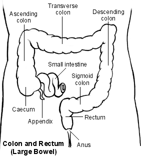
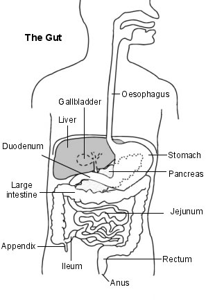
CT colonography is known in full as computed tomography colonography (sometimes the word computerised is used instead of computed). It is a test that uses a CT scanner to produce pictures of the inside of the colon and rectum (the colon, or large intestine, is the last part of the intestines or guts, the rectum is the passage between the colon and the anus).
Colon and Rectum
Digestive system
CT colonography can provide information about the colon that would usually only be obtainable by doing a colonoscopy. Colonoscopy is a test where a flexible tube is inserted into the rectum and through the colon. See the separate leaflet called Colonoscopy for more details.
CT colonography is less invasive than a colonoscopy because the test does not involve inserting a tube all the way around the colon. CT colonography is sometimes referred to as virtual colonoscopy.
What is CT colonography used for?
The major reason for doing CT colonography is to look for polyps or cancers in the colon or rectum. Polyps are small growths on the inside of your bowel. They are usually harmless but some polyps can develop into bowel cancer.
CT colonography can be used if you have symptoms such as changes in your bowel habit, weight loss or blood in your faeces (stools). It can also be used to screen people who are at risk of developing bowel cancer.
CT colonography is often used in people who are too frail to have a colonoscopy, or if there are other reasons why a colonoscopy would not be suitable.
How does CT colonography work?
The CT scanner looks like a giant thick ring. Within the wall of the scanner there is an X-ray source. Opposite the X-ray source, on the other side of the ring, are X-ray detectors. You lie on a couch which slides into the centre of the ring until the part of the body to be scanned is within the ring. The X-ray machine within the ring rotates around your body. As it rotates around, the X-ray machine emits thin beams of X-rays through your body, which are detected by the X-ray detectors.
The detectors detect the strength of the X-ray beam that has passed through your body. The more dense the tissue, the less X-rays pass through. The X-ray detectors feed this information into a computer. Different types of tissue with different densities show up as a picture on the computer monitor, in different colours or shades of grey. So, in effect, a picture is created by the computer of a cross-section (slice) of a thin section of your body.
As the couch moves slowly through the ring the X-ray beam passes through the next section of your body. So, several cross-sectional pictures (slices) of the part of your body being investigated are made by the computer. Newer scanners can even produce 3-dimensional pictures from the data received from the various slices of the part of the body being scanned.
What happens during CT colonography?
The way in which this test is done varies between different hospitals and may also vary according to why you are having the test.
You may be given an injection of a muscle relaxant to help relax the muscles of your bowel wall. You may also be given an injection of contrast agent at the same time, depending on the reason for the test.
The test begins by positioning you on the CT examination table, usually lying flat on your back, or possibly on your side or on your stomach. Straps and pillows may be used to help you maintain the correct position during the scan.
A very small, flexible tube will be passed a small way into your rectum to allow gas (carbon dioxide) to be gently pumped into the colon. Sometimes an electronic pump is used to put gas into the colon. The gas helps to open (distend) the colon as much as possible which gets rid of any folds or wrinkles that might hide polyps or growths.
When this happens, you may briefly feel pains similar to trapped wind. You may also have the urge to go to the toilet but, because your colon is empty, this won't happen. You may pass wind, but there is no need to feel embarrassed, as the staff expect this may happen.
Next, the table will move through the scanner. You may be asked to hold your breath for about 15 seconds before turning over and lying on your back or side. As you move through the scanner the pictures will be taken.
Once the scan is done, the tube is removed and you will be allowed to get down from the table.
Is CT colonography painful?
The CT scan itself is painless. You cannot see or feel X-rays. You will be asked to stay as still as possible, as otherwise the scan pictures may be blurred. The scan can take about 5-30 minutes, depending on which part (or parts) of the body is scanned.
What should I do to prepare for CT colonography?
You should be given instructions by the CT department detailing what to do. As a general rule, for your doctor to be able to see your bowel clearly, you may need to follow a special diet for a few days before the procedure and take a laxative to empty your bowel.
You may be asked to swallow a fluid called a dye (contrast agent) around two days before the test. This fluid will help to show your bowel more accurately on the scan. If you need an injection of contrast agent it may be necessary to stop certain medicines before the procedure.This may apply to people taking metformin, a medicine used to treat diabetes. If you are taking this medication, your doctor should give you instructions about what to do.
You may be asked not to eat or drink for a few hours before your scan.
What can I expect after CT colonography?
You will usually be able to return to your normal activities as soon as the scan is over. You may feel bloated or find you pass wind for a time after the test. This should usually settle fairly quickly.
If you have had muscle relaxants or an injection of dye (contrast agent), you may need to wait for a short time before driving. You may want to arrange for someone to drive you home after the scan.
If you are breastfeeding, you may not need to express and discard your milk for 24 hours after the scan, as many research papers suggest it is safe to continue breastfeeding as normal. Ask your doctor or radiographer for specific advice.
Are there any side-effects or complications from CT colonography?
Pregnant women, if possible, should not have CT colonography, as there is a small risk that X-rays may cause an abnormality to the unborn child. Tell your doctor if you are, or think you may be, pregnant.
The laxatives used to prepare your bowel can cause diarrhoea and may make you feel sick and bloated. Occasionally, some people sense a warm feeling or get a metallic taste in their mouth after having a contrast injection. This usually lasts only a minute or two. If you have had muscle relaxants, these can temporarily blur your vision or make you feel dizzy.
Rarely, some people have an allergic reaction to the dye (contrast agent) which is sometimes used. This can be treated immediately. Very rarely, the dye may cause some kidney damage, most commonly in people who already have kidney problems.
There is a very small chance that your colon may be damaged during the procedure. This can lead to bleeding and infection, which may need treatment with medicines or surgery.
Risks of X-ray radiation used in CT scans
CT scans use X-rays, which are a type of radiation. Exposure to large doses of radiation is linked to developing cancer or leukaemia - often many years later.
The dose of X-ray radiation needed for a CT scan is much more than for a single X-ray picture, but is still generally quite a low dose. The risk of harm from the dose of radiation used in CT scanning is thought to be very small, but it is not totally without risk.
As a rule, the higher the dose of radiation, the greater the risk. So, for example, the larger the part of the body scanned, the greater the radiation dose. And, repeat CT scans over time cause an overall increase of dose. Also, the younger you are when you have a CT scan, the greater the lifetime risk of developing cancer or leukaemia.
In general, the risk of developing cancer or leukaemia following a CT scan is small. In many situations, the benefit of a CT scan greatly outweighs the risk. However the radiation doses from CT scans are kept as low as possible in order to minimise any risk.
Patient picks for Imaging
Tests and investigations
Radionuclide scan
A radionuclide scan is a way of imaging bones, organs and other parts of the body by using a small dose of a radioactive chemical. There are different types of radionuclide chemical. The one used depends on which organ or part of the body is to be scanned. Note: the information below is a general guide only. The arrangements, and the way tests are performed, may vary between different hospitals. Always follow the instructions given by your doctor or local hospital.
by Dr Rachel Hudson, MRCGP

Tests and investigations
Barium tests
Barium tests are used to help see the outline of various parts of the gut (gastrointestinal tract). These include the gullet (oesophagus), stomach, upper gut (small intestines) and colon (large intestine). Barium X-ray tests are done less commonly these days. Today we usually look into the gut with a flexible telescope (endoscopy or colonoscopy) . However, there is still a place for barium tests to help assess various problems of the gut. Note: the information below is a general guide only. The arrangements, and the way tests are performed, may vary between different hospitals. Always follow the instructions given by your doctor or local hospital.
by Dr Rosalyn Adleman, MRCGP
Frequently asked questions
Does a CT colonography show other organs?
CT colonography is primarily focused on imaging the colon and rectum. However, the CT scan does pass through other body parts in the abdomen and pelvis. While not the main purpose, some other organs might be incidentally visible on the images.
Why is CT colonography sometimes called a 'virtual colonoscopy'?
CT colonography is referred to as a 'virtual colonoscopy' because it produces detailed images of the inside of the colon and rectum, similar to what a traditional colonoscopy would show, but without requiring the insertion of a long, flexible tube all the way through the colon. It uses a CT scanner to create these 'virtual' views.
Can I have a CT colonography if I have a pacemaker or other medical implants?
The article does not specifically mention pacemakers or other medical implants in relation to CT colonography. CT scans use X-rays, not strong magnetic fields like MRI, so medical implants are generally not an issue. However, you should always inform your doctor and the CT department about any medical devices you have.
What happens if polyps or other abnormalities are found during the CT colonography?
The article states that CT colonography is used to look for polyps or cancers. If such abnormalities are found, the next steps are not detailed in this article. Typically, this would involve further consultation with your doctor to discuss findings and potential follow-up procedures, such as a traditional colonoscopy for removal or biopsy.
How accurate is CT colonography compared to a traditional colonoscopy in detecting problems?
The article mentions that CT colonography can provide information about the colon that would usually only be obtainable by doing a colonoscopy, and it is used to look for polyps or cancers. However, it does not provide a direct comparison of the accuracy or detection rates between CT colonography and traditional colonoscopy.
Further reading and references
- Computed tomographic colonography (virtual colonoscopy); NICE Interventional Procedures Guidance, June 2005
- Halligan S; CT colonography for investigation of patients with symptoms potentially suggestive of colorectal cancer: a review of the UK SIGGAR trials. Br J Radiol. 2013 Jun;86(1026):20130137. doi: 10.1259/bjr.20130137. Epub 2013 Apr 8.
- Plumb AA, Halligan S, Nickerson C, et al; Use of CT colonography in the English Bowel Cancer Screening Programme. Gut. 2014 Jun;63(6):964-73. doi: 10.1136/gutjnl-2013-304697. Epub 2013 Aug 16.
- Sali L, Regge D; CT colonography for population screening of colorectal cancer: hints from European trials. Br J Radiol. 2016 Dec;89(1068):20160517. doi: 10.1259/bjr.20160517. Epub 2016 Sep 14.
- Cova MA, Stacul F, Quaranta R, et al; Radiological contrast media in the breastfeeding woman: a position paper of the Italian Society of Radiology (SIRM), the Italian Society of Paediatrics (SIP), the Italian Society of Neonatology (SIN) and the Task Force on Breastfeeding, Ministry of Health, Italy. Eur Radiol. 2014 Aug;24(8):2012-22. doi: 10.1007/s00330-014-3198-6. Epub 2014 May 17.
About the author
Updated by

Dr Rachel Hudson, MRCGP
General Practitioner and Medical Author
MBChB, MRCGP (2008), BSc (Medical Science), DFSRH, DRCOG, DCH
Dr Rachel Hudson, is an NHS GP working in the North West of England.
Authored by

Dr Tim Kenny, MRCGP
BMedSci (Hons), MB BS, MRCGP, DCH
About the reviewerView full bio

Dr Krishna Vakharia, MRCGP
Chief Medical Officer for Health, Optum UK
MBChB, MRCGP(2013), BMedSci (hons), DFSRH, DRCOG, PGDipDerm (Distn)
Dr Krishna Vakharia is an NHS GP. She is also a regular examiner for the postgraduate Diploma in Practical Dermatology at Cardiff University as well as being the Chief Medical Officer for health at Optum UK.
Article history
Article also available in English, German, Spanish, French, Italian, Portuguese, Hindi, Hebrew, Arabic, and Swedish.
Next review due: 23 Feb 2028
8 Mar 2023 | Latest version
19 Jul 2012 | Originally published
Authored by:
Dr Tim Kenny, MRCGP
Join the conversation
Share your thoughts or experiences with others in our patient communities.

Ask, share, connect.
Browse discussions, ask questions, and share experiences across hundreds of health topics.

Feeling unwell?
Assess your symptoms online for free
Sign up to the Patient newsletter
Your weekly dose of clear, trustworthy health advice - written to help you feel informed, confident and in control.
By subscribing you accept our Privacy Policy. You can unsubscribe at any time. We never sell your data.