Pression veineuse jugulaire
Revu par Dr Philippa Vincent, MRCGPDernière mise à jour par Dr Toni Hazell, FRCGPDernière mise à jour 30 Jul 2026
Respecte les directives éditoriales
- TéléchargerTélécharger
- Partager
- Language
- Discussion
- Ask Patient
- Version audio
- Ajouter aux sources préférées sur Google
Professionnels de la santé
Les articles de référence professionnelle sont conçus pour être utilisés par les professionnels de la santé. Ils sont rédigés par des médecins britanniques et basés sur des preuves de recherche, des directives britanniques et européennes. Vous pouvez trouver l'un de nos articles de santé plus utile.
What is jugular venous pressure?
Jugular venous pressure (JVP) provides an indirect measure of central venous pressure. The internal jugular vein connects to the right atrium without any intervening valves - thus acting as a column for the blood in the right atrium. The JVP consists of certain waveforms and abnormalities of these can help to diagnose certain conditions.1 Unfortunately, detection of these abnormalities and even the JVP itself, can be difficult and has also been superseded by other diagnostic methods.
How to examine jugular venous pressure1
Use the right internal jugular vein (IJV).
The patient should be at a 45° angle.
The patient's head should be turned slightly to the left.
If possible, have a tangential light source that shines obliquely from the left.
Locate the surface markings of the IJV - this runs from the medial end of the clavicle to the ear lobe, under the medial aspect of the sternocleidomastoid.
Locate the JVP - look for the double waveform pulsation (palpating the contralateral carotid pulse will help).
Measure the level of the JVP by measuring the vertical distance between the sternal angle and the top of the JVP. Measure the height - usually less than 3 cm.
Waveforms of jugular venous pressure
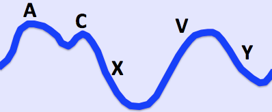
Jugular venous pulse
Informations importantes |
|---|
Waves1 a - presystolic; produced by right atrial contraction. c - bulging of the tricuspid valve into the right atrium during ventricular systole (isovolumic phase). v - occurs in late systole; increased blood in the right atrium from venous return. Descents x - a combination of atrial relaxation, downward movement of the tricuspid valve and ventricular systole. y - the tricuspid valve opens and blood flows into the right ventricle. |
The a and v waves can be identified by timing the double waveform with the opposite carotid pulse. The a wave will occur just before the pulse and the v wave occurs towards the end of the pulse. Distinguishing the c wave, x and y descents is an almost impossible task.
How to differentiate a jugular venous pulse from the carotid pulse
The jugular venous pulse is:
Not palpable.
Obliterated by pressure.
Characterised by a double waveform.
Variable with respiration - it decreases with inspiration.
Enhanced by the hepatojugular reflux (see below).
Hepatojugular reflux (abdominojugular reflux sign)2
This can help to confirm that the pulsation is caused by the JVP.
In the classic test for hepatojugular reflux, firm pressure is applied to the right upper quadrant using the palm of the hand. It has been realised that pressure anywhere over the abdomen will produce the same result (abdominojugular reflux sign). Pressure over the peri-umbilical region is the usual method and may be more appropriate in patients with a tender liver.
A transient increase in the JVP will be seen in normal patients.
There may be a delayed recovery back to baseline which is more marked in right ventricular failure.
Raised jugular venous pressure causes
Insuffisance cardiaque.
Constrictive pericarditis (JVP increases on inspiration - called Kussmaul's sign).
.
Fluid overload - eg, renal disease.
Superior vena cava obstruction (no pulsation).
Jugular venous pressure abnormalities13
Abnormalities of the a wave
It disappears in atrial fibrillation.
Large a waves occur in any cause of right ventricular hypertrophy (pulmonary hypertension and pulmonary stenosis) and tricuspid stenosis.
Extra large a waves (called cannon waves) in complete heart block and ventricular tachycardia.
Prominent v waves
Tricuspid regurgitation - called cv or v waves and occurring at the same time as systole (a combination of v wave and loss of x descent); there may be earlobe movement.
Slow y descent
Tricuspid stenosis.
Right atrial myxoma.
Steep y descent
Insuffisance ventriculaire droite.
Péricardite constrictive.
Tricuspid regurgitation.
(The last two conditions have a rapid rise and fall of the JVP - called Friedreich's sign.)
Prognostic use of jugular venous pressure
Elevated JVP in patients with heart failure is associated with an increased risk of hospital admission, death and subsequent hospitalisation for heart failure.4 Therefore, appreciation of this sign can be clinically helpful.
Mises à jour exclusives pour les professionnels de la santé
Restez informé des dernières mises à jour cliniques, des perspectives professionnelles et des conseils fondés sur des preuves. La newsletter Patient Pro sélectionne des contenus essentiels pour les professionnels de santé—livrés directement dans votre boîte de réception.
En vous abonnant, vous acceptez notre Politique de confidentialité. Vous pouvez vous désabonner à tout moment. Nous ne vendons jamais vos données.
Lectures complémentaires et références
- Chua Chiaco JM, Parikh NI, Fergusson DJ; The jugular venous pressure revisited. Cleve Clin J Med. 2013 Oct;80(10):638-44. doi: 10.3949/ccjm.80a.13039.
- Brostoff JM; Re-examining examination: misconceptions in clinical medicine. J R Soc Med. 2009 Jan;102(1):11-5. doi: 10.1258/jrsm.2008.080330.
- Witteles R; Neck Veins & Wave Forms, Stanford Medicine 25
- Drazner MH, Rame JE, Stevenson LW, et al; Prognostic importance of elevated jugular venous pressure and a third heart sound in patients with heart failure. N Engl J Med. 2001 Aug 23;345(8):574-81.
À propos de l'auteurVoir la biographie complète
Mis à jour par

Dr Toni Hazell, FRCGP
MBBS, BSc, FRCGP, DFSRH, Dip GU med, DRCOG, DCH (London, UK, 2000)
Le Dr Toni Hazell a obtenu son diplôme de l'École de médecine de l'hôpital St. Mary et a effectué son VTS à l'hôpital Northwick Park.
À propos du critiqueVoir la biographie complète

Dr Philippa Vincent, MRCGP
Médecin généraliste, Auteur médical
MB BS, Bsc, MRCGP (2000), DCH, DFSRH, DRCOG
Dr Philippa Vincent est un médecin généraliste du NHS travaillant dans le nord de Londres.
Accès simple à des traitements fiables et à des services de santé en ligne.
Common conditions
Get help with common conditions through the NHS, without a GP appointment.




Plus dans l'histoire et l'examen
- Vertige positionnel paroxystique bénin
- Masse mammaire et examen des seins
- Étouffement et obstruction des voies respiratoires par un corps étranger
- Toux chronique chez les enfants
- Douleur chronique
- Otite moyenne suppurée chronique
- Vision des couleurs et ses troubles
- Coma
- Diplopie et lésions des nerfs crâniens III, IV et VI
- Test de tolérance à l'exercice
- Syndrome de Foster Kennedy
- Infections fongiques des ongles
- Anomalies de la démarche chez les enfants
- Hoquet
- Score international des symptômes de la prostate
- Douleur lombaire
- Tachycardies à complexes étroits
- Obésité chez les enfants
- Tremblement
- Testicules non descendus