IRM
Revu par des pairs Dr Colin Tidy, MRCGPDernière mise à jour par Dr Toni Hazell, FRCGPDernière mise à jour 7 août 2023
Respecte les directives éditoriales
- TéléchargerTélécharger
- Partager
- Language
- Discussion
- Version audio
- Ajouter aux sources préférées sur Google
An MRI scan is a safe and painless test that can provide detailed pictures of organs and other structures inside your body.
Remarque: the information below is a general guide only. The arrangements (and the way tests are performed) may vary between different hospitals. Always follow the instructions given by your doctor or local hospital. These are usually included with your appointment letter.
En un coup d'œil
Une IRM utilise un champ magnétique puissant et des ondes radio pour créer des images détaillées de l'intérieur de votre corps.
Il peut montrer différents tissus, organes et autres structures, aidant au diagnostic de diverses conditions.
Vous êtes allongé sur un canapé à l'intérieur d'un scanner en forme de tunnel, et vous devez rester immobile pendant le scan de 15 à 40 minutes.
Le scan est indolore mais peut être bruyant, donc on vous proposera des écouteurs ou des bouchons d'oreilles.
Informez votre médecin si vous avez peur des espaces clos ou si vous avez des implants métalliques en raison de l'aimant puissant.
Il est généralement déconseillé aux femmes enceintes de passer une IRM sauf en cas d'urgence, car les effets à long terme sur un bébé sont inconnus.
Qu'est-ce qu'une IRM ?
MRI stands for magnetic resonance imaging. An MRI scan uses a strong magnetic field and radio waves to create a detailed image on a computer screen. It can show different types of tissue, organs and other structures inside your body.
This image is an MRI scan of a brain. The person's eyeballs can be seen at the top of the picture.
IRM
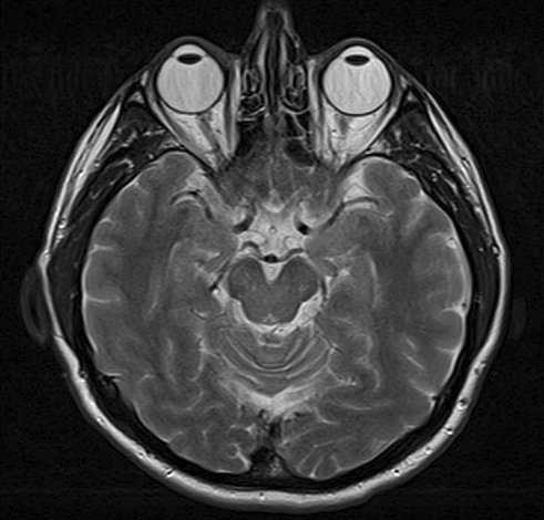
© By Ptrump (Own work) via Wikimedia Commons
How does an MRI scan work?
Your body contains millions of hydrogen atoms. When you are in an MRI scanner:
A strong magnetic field aligns particles called protons which are within the hydrogen atoms. All the protons line up in parallel to the magnetic field, like tiny magnets. (Normally the millions of protons all lie in random directions.)
Then short bursts of radio waves are sent from the scanner into your body. The radio waves knock the protons from their position.
When the burst of radio waves stop, the protons realign back into place. As they do so they emit radio signals. The protons in different tissues of the body realign at different speeds. Therefore, the signal emitted from different body tissues varies. So, for example, softer tissues can be distinguished from harder tissues on the basis of the signals sent.
These signals are detected by a receiving device in the scanner.
The receiving device transmits the signals to a computer. The computer creates a picture based on the radio signals emitted from the body.
What does an MRI scan involve?
The MRI scanner is like a tunnel about 1.5 metres long, surrounded by a large circular magnet. You lie on a couch which then slides into the scanner. A 'receiving device', like an aerial, is placed behind, or around, the part of the body being examined. This detects the tiny radio signals emitted from your body. When each 'picture' is being taken you need to keep still for a few minutes, otherwise the scan picture may be blurred.
MRI scanner
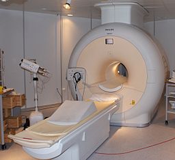
© By Jan Ainali (own work) via Wikimedia Commons.
The scan itself is painless. The whole procedure can take 15-40 minutes. It may be a little uncomfortable lying still on the couch for this time. Small children may need a general anaesthetic to keep them still long enough for the pictures to be taken. Where you lie is quite enclosed and some people may find this very unsettling.
If you have a fear of confined spaces (claustrophobia) you should discuss this with your doctor before you go for the scan. Some parts of the country have access to 'open' scan machines. However, they are not widely available and there is usually a longer wait for an 'open' scanner than for a closed scanner.
In some cases, an injection of a special contrast dye is given into the bloodstream via a vein on the arm. This helps to give clearer pictures of certain tissues or organs being examined.
The radiographer sits in the control room next to the scanner and observes through the window. However, you can talk to them, usually via an intercom, and you will be observed at all times on a monitor.
The scanner is noisy so you will be given some headphones or earplugs to protect your ears from the noise. Quite often you can listen to the radio or music through the headphones.
What is an MRI scan used for?
An MRI scan can create clear pictures of most parts of the body. So, it is useful for all sorts of reasons where other tests (such as les radiographies) do not give enough information required.
It is commonly used to obtain detailed pictures of the brain and spinal cord, to detect abnormalities and tumours. Even torn ligaments around joints can be detected by an MRI scan. So it is being used more and more following sports injuries.
Cerveau
MRI is the first-choice investigation for tumeurs cérébrales, as it produces clearer images than computerised tomography (CT) and shows hard-to-reach areas of the brain. There is clear contrast between grey and white matter parts of the brain and this makes MRI the best choice for many other conditions, including sclérose en plaques, AVC, Maladie d'Alzheimer et épilepsie.
Musculoskeletal system
Here, MRI is used to look at the spine - to assess joint disease, pressure on nerves and soft tissue tumours.
Gastrointestinal system
MRI allows non-invasive assessment of la maladie inflammatoire de l'intestin et bowel tumours. It can also look at problems in the liver and pancreas.
Blood vessels and the heart
This is called magnetic resonance angiography (MRA) and it generates pictures of the arteries to look for abnormal narrowing or vessel wall dilatations (those at risk of bursting). MRA is often used to evaluate the arteries of the neck and brain, the thoracic and abdominal aorta, the renal arteries, and the legs. It might also be used to assess congenital heart disease.
Preparing for an MRI scan
Your local hospital should give you information about what is required before you come for the scan.
The MRI scanner uses an extremely strong magnet, so people with certain types of medical implant cannot be scanned. This is because the magnet can potentially move medical devices with metal in them, or affect their function.
Therefore, before you enter the scanning area you should be asked if you have any medical devices in your body. You may have to fill in a safety questionnaire that asks about things that may contain metal. The following is not a definitive list but may help to remind you of the type of things radiographers need to know about:
Internal (implanted) defibrillator or pacemaker.
Ear (cochlear) implant.
Surgical clips such as those used on brain aneurysms.
Artificial heart valves.
Implanted medicine infusion ports.
Artificial limbs or metallic joints.
Implanted nerve stimulators.
Pins, screws, plates, stents or surgical staples.
It is also important to tell the radiographer if you have ever had any metal fragments lodged in your eyes or your body. In some cases you may need an X-ray before an MRI scan, to make sure you are safe to enter the scanner.
Side-effects of an MRI scan
MRI scans are painless and thought to be safe. MRI scans do not use X-rays so the possible concerns associated with X-ray pictures and Scans CT (which use X-rays) are not associated with MRI scans.
However:
Rarely, some people have reactions to the contrast agent which is sometimes used.
Pregnant women are usually advised not to have an MRI scan unless it is urgent. Although the scan is thought to be safe, the long-term effects of strong magnetic fields on a developing baby are not yet known.
What to expect after the MRI scan
There are no after effects from the scan. You can return to your normal activities as soon as the scan is over. The scan pictures are studied by a specialist who interprets them - a radiologist - who sends a report to the doctor who requested the scan.
It is usual to have to wait for at least two weeks before hearing about your results. If there are any urgent findings, the specialist will be informed as soon as possible. The result will go back to the person who requested the scan. So, if a hospital consultant requests a scan, you will get the result at your next appointment with them. If you are concerned, you could ring their secretary, but do not contact the GP for the result as they will not have it. If the GP has requested an MRI, you will get the result from them.
Sélections des patients pour Imagerie

Tests et investigations
Radiographie
Les radiographies montrent les os et certains autres tissus.
par Dr Hayley Willacy, FRCGP

Tests et investigations
Scan TEP
Un scanner TEP crée des images qui montrent où les cellules sont particulièrement actives dans le corps. Il est le plus souvent utilisé pour diagnostiquer et évaluer le cancer. Remarque : les informations ci-dessous sont uniquement un guide général. Les dispositions et la manière dont les tests sont effectués peuvent varier entre différents hôpitaux. Suivez toujours les instructions données par votre médecin ou votre hôpital local.
par Dr Rosalyn Adleman, MRCGP
Questions fréquemment posées
Puis-je conduire une voiture immédiatement après avoir passé une IRM ?
Oui, il n'y a pas d'effets secondaires après une IRM, vous pouvez donc généralement reprendre vos activités normales, y compris conduire, dès que l'examen est terminé.
Combien de temps faudra-t-il pour obtenir les résultats de mon IRM ?
Il faut généralement au moins deux semaines pour recevoir les résultats de votre IRM. Cependant, s'il y a des résultats urgents, le spécialiste informera le médecin demandeur dès que possible. Les résultats seront envoyés au médecin qui a demandé l'examen ; s'il s'agissait d'un consultant hospitalier, vous les recevrez lors de votre prochain rendez-vous avec lui. Si votre médecin généraliste a demandé l'IRM, il vous fournira les résultats.
Que dois-je faire si j'ai peur des espaces clos ?
Si vous avez peur des espaces confinés (claustrophobie), vous devriez en discuter avec votre médecin avant votre examen. Bien que des machines d'imagerie 'ouvertes' existent, elles ne sont pas largement disponibles et impliquent généralement un temps d'attente plus long par rapport aux scanners fermés plus courants.
Pourquoi me donnerait-on une injection lors d'une IRM ?
Dans certains cas, un colorant de contraste spécial est injecté dans votre circulation sanguine par une veine de votre bras. Ce colorant aide à produire des images plus claires des tissus ou organes spécifiques qui sont examinés pendant le scan.
Serai-je seul pendant l'examen IRM ?
Non, vous ne serez pas seul. Le radiographe sera dans une salle de contrôle à côté du scanner, vous observant à travers une fenêtre et sur un moniteur en permanence. Vous pouvez communiquer avec lui, généralement via un système d'interphone.
Lectures complémentaires et références
- IRM; Cancer Research UK; 2022
À propos de l'auteurVoir la biographie complète

Dr Toni Hazell, FRCGP
MBBS, BSc, FRCGP, DFSRH, Dip GU med, DRCOG, DCH (London, UK, 2000)
Le Dr Toni Hazell a obtenu son diplôme de l'École de médecine de l'hôpital St. Mary et a effectué son VTS à l'hôpital Northwick Park.
À propos du critiqueVoir la biographie complète

Dr Colin Tidy, MRCGP
Médecin généraliste, Auteur médical
MBBS, MRCGP, MRCP (Paediatrics), DCH
Le Dr Colin Tidy est un médecin du NHS, basé dans l'Oxfordshire.
Historique de l'article
Les informations sur cette page sont rédigées et examinées par des cliniciens qualifiés.
Article également disponible en Anglais, Allemand, Espagnol, Français, Italien, Portugais, Hindi, Hébreu, Arabe, and Suédois.
Prochaine révision prévue : 5 août 2028
7 août 2023 | Dernière version

Demandez, partagez, connectez-vous.
Parcourez les discussions, posez des questions et partagez vos expériences sur des centaines de sujets de santé.

Vous ne vous sentez pas bien ?
Évaluez vos symptômes en ligne gratuitement
Inscrivez-vous à la newsletter Patient
Votre dose hebdomadaire de conseils de santé clairs et fiables - rédigés pour vous aider à vous sentir informé, confiant et maître de la situation.
En vous abonnant, vous acceptez notre Politique de confidentialité. Vous pouvez vous désabonner à tout moment. Nous ne vendons jamais vos données.
Plus dans les tests et enquêtes
- Tests d'anticorps et d'antigènes
- Audiologie
- Biopsie
- Tests de coagulation sanguine
- Analyses de sang pour détecter l'inflammation
- Scintigraphie osseuse
- Gènes BRCA
- Angiographie cérébrale
- Scan CT
- Cystoscopie
- Électroencéphalographe
- Échographie endoscopique
- Aspiration à l'aiguille fine
- Urographie intraveineuse
- IRMCP
- Test auditif néonatal
- Protéinurie
- Test sanguin de routine pour la fonction rénale
- Spirométrie
- Échographie