Urographie intraveineuse
Pyelography
Revu par Dr Hayley Willacy, FRCGP Dernière mise à jour par Dr Doug McKechnie, MRCGPLast updated 31 oct. 2023
Respecte les directives éditoriales
- TéléchargerTélécharger
- Partager
- Language
- Discussion
- Version audio
- Add to preferred sources on Google
Urographie intraveineuse is a test that uses X-rays and a special dye to help assess your kidneys, ureters, bladder and urethra.
Remarque: les informations ci-dessous ne sont qu'un guide général. Les dispositions et la manière dont les tests sont effectués peuvent varier d'un hôpital à l'autre. Suivez toujours les instructions données par votre médecin ou votre hôpital local.
At a glance
Intravenous urography is an X-ray procedure assessing your urinary tract using an injected dye.
It helps detect kidney stones, causes of recurring urine infections, and blood in urine.
You must inform your doctor of any allergies, especially to contrast dyes like iodine.
The procedure usually takes 30-60 minutes and involves taking several X-ray pictures.
Common side-effects include a warm feeling or metallic taste in your mouth.
Pregnant women should avoid this test due to a small risk to the unborn child.
Dans cet article:
Video picks for Tests d'urine et de vessie
Continuez à lire ci-dessous
What is intravenous urography?
Cross-section diagram of the urinary tract describing how an intravenous urography is performed
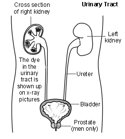
Intravenous urography (also known as intravenous pyelography) is an X-ray procedure which is used to assess problems in your kidneys, ureters, bladder and urethra. These structures make up your urinary tract. The ureters are tubes which go from each kidney to your bladder. The urethra is the tube from your bladder that passes out urine.
The urinary tract does not show up well on ordinary X-ray pictures. However, with intravenous urography a contrast dye is injected into a vein (an 'intravenous' injection). The dye travels in your bloodstream, concentrates in your kidneys, and is passed out into your ureters with urine made by your kidneys.
The dye blocks X-rays so the structure of your kidneys, ureters and bladder shows up clearly as white on X-ray pictures.
The X-ray pictures produced are called an intravenous urogram (IVU) but can also be called an intravenous pyelogram (IVP).
What is intravenous urography used for?
Retour au sommaireIntravenous urography can help to assess a range of problems. For example:
Kidney stones. A stone in a kidney or in the tube which goes from a kidney to the bladder (the ureter) will normally show up quite clearly.
Urine infections. If you have infections of your bladder or kidney which come back (recur), an IVU may help to find if you have a blockage or other abnormality of your urinary tract.
Sang dans les urines. This can be due to various causes such as infection, inflammation and tumours of the kidney. An IVU may help to clarify the cause.
Obstruction or damage to any part of the urinary tract can often be seen on an IVU.
Intravenous urography has mostly been replaced by CT urography and IRM urography scans, as these tend to give better images of the bladder, ureters, and kidneys. However, intravenous urography is still sometimes used, especially if CT or MRI scans are not available.
Continuez à lire ci-dessous
How to prepare for intravenous urography
Retour au sommaireYour kidneys have to be able to filter the dye. Therefore, it is seldom performed if you have kidney failure. Before the procedure you may need a blood test to check that you do not have kidney failure.
Tell your doctor if you have any allergies, especially allergy to contrast dyes such as iodine.
You may be asked not to eat for several hours before the procedure. This ensures that your gut (intestines) is empty of food, which makes the X-ray pictures clearer.
You may be given some laxatives to take for a day or so before the procedure. The aim of this is to clear the intestines, which will make the X-ray pictures clearer.
You may be asked to sign a consent form to confirm that you understand the procedure.
You will need to remove any metal objects or jewellery that might interfere with the X-ray pictures.
If you have diabetes and take a medicine called metformin you may need to stop the metformin for two days prior to the procedure. This is because the combination of metformin and contrast dye may affect the kidneys. (You should discuss this, and how to manage your diabetes over this period, in more detail with your doctor.)
Note de l'éditeur
Dr Krishna Vakharia, 1st November 2024
If you are having a procedure with contrast it is also important to let the department know if you have kidney disease or have had a kidney transplant. Let them know if you are under a kidney specialist or a urologist. You may also be asked to stop certain medications such as some used for high blood pressure before a procedure with contrast. Your doctor or scan department will discuss this with you if this is something you need to do.
How is intravenous urography done
Retour au sommaireYou will be asked to wear a hospital gown and to lie on a couch.
Contrast dye is then injected into a vein in your hand or arm. This may sting a little. The dye then starts to filter through the kidneys into the tubes which go from each kidney to the bladder (the ureters).
A series of X-ray pictures is then taken over your tummy (abdomen), usually every 5-10 minutes for up to 60 minutes.
You stay on the couch between each X-ray picture; however, you may be asked to get up to empty your bladder before the final X-ray picture is taken.
The procedure usually takes about 30-60 minutes. Some pictures, however, may be taken hours later in certain circumstances.
You should be able to go home as soon as the procedure is finished. You can eat normally straight afterwards.
Continuez à lire ci-dessous
Side-effects from intravenous urography
Retour au sommaireAn intravenous urogram involves exposure to X-rays, a type of radiation. This can increase the risk of developing cancer in the future, although the additional risk is low. The amount of radiation from an intravenous urogram is about the same as the amount of radiation that you would be exposed to in a year's worth of normal life.
Common side-effects include:
A flushing or warm feeling at the injection site when the dye is being injected.
A metallic taste in the mouth.
Uncommon side-effects include:
An allergic reaction to the dye.
Acute kidney injury.
An allergic reaction to the dye occurs in a small number of cases. Symptoms may be mild - for example, an itchy skin rash and some mild swelling of the lips. More severe symptoms are extremely rare - for example, breathing difficulties and collapse due to pression artérielle basse.
Who should not have intravenous urography?
Retour au sommairePregnant women, if possible, should not have any X-ray tests, as there is a small risk that X-rays may cause an abnormality to the unborn child. This is why women are asked before having an X-ray if they are, or might be, pregnant.
Patient picks for Tests d'urine et de vessie
Tests et investigations
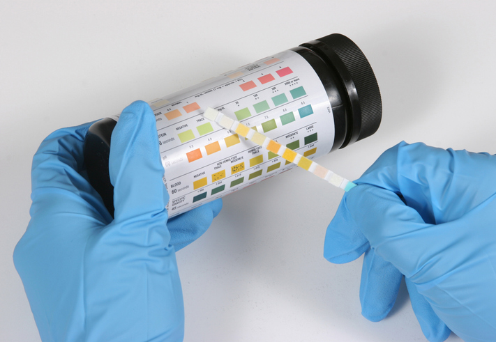
Test de bandelette urinaire
A urine dipstick test tests the urine using a special strip of paper that is dipped into a sample of urine. The result is available almost immediately. It is sometimes called a rapid urine test.
par Dr Philippa Vincent, MRCGP

Tests et investigations
Test urodynamique
Les tests urodynamiques vérifient le remplissage et la vidange de votre vessie et aident à rechercher la cause de toute incontinence urinaire que vous pourriez avoir. Remarque : les informations ci-dessous sont uniquement un guide général. Les modalités et la manière dont les tests sont effectués peuvent varier d’un hôpital à l’autre. Suivez toujours les instructions données par votre médecin ou votre hôpital local.
by Dr Hayley Willacy, FRCGP
Questions fréquemment posées
What is the difference between an intravenous urogram (IVU) and an intravenous pyelogram (IVP)?
An intravenous urogram (IVU) and an intravenous pyelogram (IVP) are different names for the same X-ray procedure. The pictures produced are called an intravenous urogram (IVU) but can also be called an intravenous pyelogram (IVP).
I have diabetes and take metformin; do I need to stop this medication before the procedure?
If you have diabetes and take metformin, you may need to stop taking it for two days before the intravenous urography. This is because the combination of metformin and the contrast dye used in the procedure could affect your kidneys. You should discuss this thoroughly with your doctor to understand how to manage your diabetes during this period.
How long will I be at the hospital for this procedure?
The intravenous urography procedure itself usually takes about 30-60 minutes to complete. You should be able to go home as soon as the procedure is finished. In some specific circumstances, some pictures might be taken hours later.
What should I expect immediately after the intravenous urography?
Immediately after the procedure, you should be able to go home. You can also eat normally straight away. You will have been lying on a couch during the X-rays, and you might have been asked to empty your bladder before the final picture.
What is the purpose of not eating and taking laxatives before the procedure?
You may be asked not to eat for several hours before the procedure, and you might be given laxatives to take for a day or so beforehand. The goal of both of these preparations is to ensure that your gut (intestines) is empty. An empty gut helps to make the X-ray pictures clearer, allowing for a better view of your urinary tract.
Are there any medical conditions that would prevent me from having an intravenous urography?
Intravenous urography is not usually performed if you have kidney failure, as your kidneys need to be able to filter the dye. Also, pregnant women, if possible, should generally avoid all X-ray tests due to a small risk to the unborn child. If you have kidney disease or have had a kidney transplant, or are under a kidney specialist or urologist, it is important to inform the department before the procedure.
Continuez à lire ci-dessous
About the authorView full bio

Dr Doug McKechnie, MRCGP
Medical Writer
MA, MBBS, MSc, DRCOG, MRCP(UK), MRCGP(2021), FHEA
Dr Doug McKechnie is an NHS GP working in London. He works full-time clinically and is also the Deputy Lead for the Clinical and Professional Practice module at University College London Medical School.
About the reviewerView full bio

Dr Hayley Willacy, FRCGP
Médecin généraliste, Auteur médical
MBChB (1992), DRCOG, DFFP, MRCOG (Part 1) MRCGP (2007), DFSRH (2013), MSc - medical education (2020)
Dr Hayley Willacy was an NHS GP working in northwest England, who retired from clinical practice in 2022 after 30 years.
Historique de l'article
Les informations sur cette page sont rédigées et examinées par des cliniciens qualifiés.
Prochaine révision prévue : 29 oct. 2028
31 oct. 2023 | Dernière version

Demandez, partagez, connectez-vous.
Parcourez les discussions, posez des questions et partagez vos expériences sur des centaines de sujets de santé.

Vous ne vous sentez pas bien ?
Évaluez vos symptômes en ligne gratuitement
Inscrivez-vous à la newsletter Patient
Votre dose hebdomadaire de conseils de santé clairs et fiables - rédigés pour vous aider à vous sentir informé, confiant et maître de la situation.
By subscribing you accept our Politique de confidentialité. Vous pouvez vous désabonner à tout moment. Nous ne vendons jamais vos données.