Maladie de Perthes
Revu par Dr Toni Hazell, FRCGPDernière mise à jour par Dr Doug McKechnie, MRCGPDernière mise à jour 14 nov. 2023
Respecte les directives éditoriales
- TéléchargerTélécharger
- Partager
- Language
- Discussion
- Version audio
- Ajouter aux sources préférées sur Google
Dans cette série :Problèmes de hancheFracture de la hancheremplacement de la hancheÉpiphysiolyse fémorale supérieureSyndrome de douleur du grand trochanter
La maladie de Perthes est une affection où la tête du fémur dans l'articulation de la hanche perd son approvisionnement en sang, ce qui endommage l'os. L'os guérit et se reforme progressivement, mais la maladie de Perthes peut entraîner des problèmes de hanche plus tard dans la vie.
En un coup d'œil
La maladie de Perthes est un trouble rare de la hanche chez l'enfant, touchant principalement les enfants âgés de 3 à 10 ans.
Elle se produit lorsque l'apport sanguin à la tête du fémur s'arrête temporairement.
Les symptômes incluent souvent une boiterie, des douleurs à la hanche, et parfois des douleurs à l'aine, à la cuisse ou au genou.
Le diagnostic implique généralement un examen physique et des radiographies, parfois avec d'autres examens d'imagerie.
Le traitement vise à aider la hanche à guérir et à maintenir correctement la tête du fémur en place.
Il peut falloir deux ans ou plus pour que la hanche guérisse.
Les jeunes enfants ont souvent de meilleurs résultats, mais il y a un risque d'arthrite plus tard dans la vie.
What is Perthes' disease?
Perthes' disease, also known as Legg-Calvé-Perthes disease, is a rare childhood hip disorder that affects the blood supply to the femoral head.
It mainly affects young children who are aged between 3 and 10 years. Each year, about 1 in 10,000 children aged less than 15 years will get Perthes' disease. It is four times more common in boys than in girls.
Perthes' disease usually only affects one hip. But both hips are affected in about 1 in 7 children who have Perthes' disease.
Symptoms of Perthes' disease
The symptoms usually develop gradually over a period of time. The first indication that a child may have Perthes' disease is when they develop a limp. The affected hip is often but not always painful. The symptoms may include:
Douleur
The affected hip is often painful and pain is also felt in the groin. The pain is also often felt in the thigh and the knee. This is not because the knee is affected but because hip pain is often felt in the knee. This is called referred pain.
Limb shortening
The leg of the affected hip may become shortened compared with the unaffected side.
Limp
Children with Perthes' disease often develop a limp, which becomes gradually worse over a few weeks.
Stiffness and a reduced range of movement
As the hip becomes more damaged, the affected hip becomes stiff. A limited range of motion of the hip develops.
Muscle wasting
Because the affected leg can't be used normally, the leg's muscles will become weaker. This causes the thigh muscles on the affected side to become wasted and so the leg looks thinner when you compare it with the other leg. This may not be obvious when both hips are affected.
What causes Perthes' disease?
Diagram detailing the hip joint
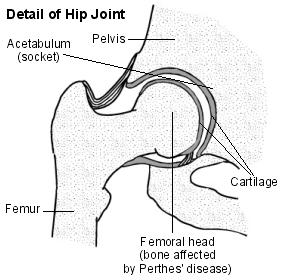
Perthes' disease occurs in a part of the hip joint called the femoral head. This is the rounded top of the thigh bone (femur) which sits inside the hip socket (acetabulum). Something happens to the small blood vessels which supply the femoral head with blood.
So, parts of the femoral head lose their blood supply. As a result, the bone cells in the affected area die, the bone softens and the bone can fracture or become distorted. This is called 'avascular necrosis'. The amount of bone damage can vary from mild to severe.
The exact cause of the blood vessel problem that occurs in the first place is not known. A child with Perthes' disease is usually otherwise well.
Over several months the blood vessels regrow, and the blood supply returns to the bone tissue. New bone tissue is then made so the femoral head reforms and regrows. This is similar to how bone reforms and regrows after any normal fracture or break to a bone. But, with Perthes' disease, it takes longer (often between two and three years).
Diagnosing Perthes' disease
The diagnosis can usually be made by a specialist, by examination of the hip, plus an radiographie. Sometimes other tests may be needed if the diagnosis is not clear or if a more detailed picture of the hip joint is needed. Possible tests may include:
An X-ray where dye is injected into the space within the hip joint (this is called an arthrogram).
Analyses de sang and a sample of fluid from the hip joint may also be needed to rule out other problems, such as an infection of the bone or the joint.
Perthes' disease treatment
The aim of treatment is to:
Promote the healing process; et
Ensure that the top of the thigh bone in the hip joint (the femoral head) remains well seated in the hip socket as it heals and regrows.
Which treatment is chosen will depend on the age of your child and how badly your child's hip is affected. Treatments may include observation, bed rest and/or using crutches, wearing a plaster cast or a special leg brace, or surgery. Your doctor will advise on the best treatment for your child.
Observation and physiotherapy
In younger children (less than 6 years old) and those with mild disease, Perthes' disease will usually heal well without any specific treatment. These children are treated by observation, often with physiotherapy or home exercises. The home exercises help to keep the hip joint mobile and in a good position in the hip socket.
Advice may also include:
To encourage swimming (to keep the hip joint active in the full range of movements); mais
To avoid activities that can lead to any heavy impact on the hip joint, such as those involving trampolines and bouncy castles.
However, any advice will be tailored to your child's needs. Your child will also need regular follow-up with their specialist to check how their femoral head is healing. Regular X-rays of their hips are usually suggested.
Médication
Painkillers may also be useful to help relieve pain. Common painkillers suggested are ibuprofène et paracétamol.
Bed rest and/or crutches
This may be needed by some children for a short time if their symptoms are bad.
Will surgery be needed?
An operation may be considered in some cases, particularly in older children or those more severely affected. Surgery can help to keep the femoral head in the right position while it heals. Surgery can also be used to improve the shape and function of the femoral head if it has not healed well.
Complications of Perthes' disease?
In many cases, the top of the thigh bone in the hip joint (the femoral head) regrows and remodels back to normal, or near-normal. The hip joint then returns to normal and is able to work as usual.
However, this can take two or more years after the condition first starts. Even after this time, there may be some stiffness remaining in the hip and there is an increased risk of l'arthrose dans la vie ultérieure.
The main problem is that the part of the thigh bone in the hip joint (the femoral head) may not re-form with a good rounded (spherical) shape, which helps it to fit well into the hip joint socket.
This can lead to permanent damage to the hip joint. This may cause stiffness of the hip joint. It can also cause arthritis of the hip joint at an earlier age than usual - for example, at around the age of 40 years.
The younger the child is when Perthes' disease develops, the better the chance of a good outcome. Children who develop Perthes' disease after about the age of 8-9 years have the highest risk of permanent hip joint problems, such as stiffness and arthritis.
The more severe the condition, the greater the risk of permanent problems with the hip joint.
Sélections des patients pour Développement

Santé des enfants
Douleurs de croissance
Les douleurs de croissance sont des douleurs, généralement dans les jambes, courantes chez les enfants. On ne sait pas ce qui cause ces douleurs, mais malgré leur nom, elles ne sont pas liées à la croissance. Elles ne sont pas graves et disparaissent d'elles-mêmes avec le temps. Un simple soulagement de la douleur suffit. Consultez un médecin si vous remarquez quelque chose d'anormal sur les jambes, ou si les douleurs font que votre enfant boîte.
par Dr Toni Hazell, FRCGP

Santé des enfants
Croissance limitée
Le nanisme est une condition médicale ou génétique qui entraîne une croissance limitée, de sorte que les personnes sont plus petites que la moyenne. Plus précisément, les personnes atteintes de nanisme ne dépassent pas 1 m 47 (4 pieds 10 pouces). Cela peut être dû à littéralement des centaines de causes différentes, donc le terme inclut en réalité de nombreuses conditions médicales. La cause et le type de nanisme influenceront donc le pronostic et les problèmes qui peuvent y être associés. La condition la plus courante causant le nanisme est l'achondroplasie.
par Dr Hayley Willacy, FRCGP
Questions fréquemment posées
Quelle est la probabilité que les deux hanches soient affectées par la maladie de Perthes ?
La maladie de Perthes affecte généralement une seule hanche. Cependant, chez environ 1 enfant sur 7 diagnostiqué avec cette condition, les deux hanches peuvent être touchées.
Combien de temps dure le processus de guérison et de régénération pour que l'os se rétablisse de la maladie de Perthes ?
Après la perte initiale de l'apport sanguin à la tête fémorale, les vaisseaux sanguins finissent par repousser, et un nouveau tissu osseux se forme. Ce processus, où la tête fémorale se reforme et repousse, prend généralement une période prolongée, souvent entre deux et trois ans.
Y a-t-il des activités spécifiques à éviter pendant le traitement de la maladie de Perthes ?
Oui, bien que la natation soit encouragée pour garder l'articulation de la hanche active, les activités qui impliquent un impact important sur l'articulation de la hanche doivent être évitées. Cela inclut des activités comme l'utilisation de trampolines et de châteaux gonflables. Les conseils seront adaptés aux besoins spécifiques de votre enfant.
Quel type de suivi est nécessaire après un diagnostic de la maladie de Perthes ?
Les enfants diagnostiqués avec la maladie de Perthes nécessiteront des rendez-vous de suivi réguliers avec leur spécialiste. Ces rendez-vous sont importants pour surveiller la guérison de la tête fémorale. Des radiographies régulières des hanches sont généralement suggérées dans le cadre de ce suivi.
Le fait d'avoir la maladie de Perthes augmente-t-il le risque d'autres problèmes de santé à l'avenir ?
Après la maladie de Perthes, il peut y avoir un risque accru de développer de l'arthrose dans la hanche affectée plus tard dans la vie. Si la tête fémorale ne se reforme pas en une forme bien arrondie, cela peut entraîner des dommages permanents et une raideur de l'articulation de la hanche, pouvant potentiellement causer de l'arthrite à un âge plus précoce, par exemple, vers 40 ans.
L'âge de mon enfant lorsqu'il contracte la maladie de Perthes affecte-t-il le résultat ?
Oui, plus un enfant est jeune lorsque la maladie de Perthes se développe, généralement meilleures sont les chances d'un bon résultat. Les enfants qui développent la condition après environ 8-9 ans ont le risque le plus élevé de rencontrer des problèmes permanents de l'articulation de la hanche, tels que la raideur et l'arthrite.
Lectures complémentaires et références
- Shah H; Perthes disease: evaluation and management. Orthop Clin North Am. 2014 Jan;45(1):87-97. doi: 10.1016/j.ocl.2013.08.005. Epub 2013 Sep 26.
- Larson AN, Sucato DJ, Herring JA, et al; A prospective multicenter study of Legg-Calve-Perthes disease: functional and radiographic outcomes of nonoperative treatment at a mean follow-up of twenty years. J Bone Joint Surg Am. 2012 Apr 4;94(7):584-92. doi: 10.2106/JBJS.J.01073.
- Boiterie aiguë chez l'enfant; NICE CKS, septembre 2025 (accès réservé au Royaume-Uni).
- Mills S, Burroughs KE; Legg Calve Perthes Disease. StatPearls Publishing. Jan 2020.
À propos de l'auteurVoir la biographie complète

Dr Doug McKechnie, MRCGP
Rédacteur Médical
MA, MBBS, MSc, DRCOG, MRCP(UK), MRCGP(2021), FHEA
Le Dr Doug McKechnie est un médecin généraliste du NHS travaillant à Londres. Il travaille à plein temps en clinique et est également le chef adjoint du module de Pratique Clinique et Professionnelle à l'École de Médecine de l'University College London.
À propos du critiqueVoir la biographie complète

Dr Toni Hazell, FRCGP
MBBS, BSc, FRCGP, DFSRH, Dip GU med, DRCOG, DCH (London, UK, 2000)
Le Dr Toni Hazell a obtenu son diplôme de l'École de médecine de l'hôpital St. Mary et a effectué son VTS à l'hôpital Northwick Park.
Historique de l'article
Les informations sur cette page sont rédigées et examinées par des cliniciens qualifiés.
Article également disponible en Anglais, Allemand, Espagnol, Français, Italien, Portugais, Hindi, Hébreu, Arabe, and Suédois.
Prochaine révision prévue : 12 nov. 2028
14 nov. 2023 | Dernière version

Demandez, partagez, connectez-vous.
Parcourez les discussions, posez des questions et partagez vos expériences sur des centaines de sujets de santé.

Vous ne vous sentez pas bien ?
Évaluez vos symptômes en ligne gratuitement
Inscrivez-vous à la newsletter Patient
Votre dose hebdomadaire de conseils de santé clairs et fiables - rédigés pour vous aider à vous sentir informé, confiant et maître de la situation.
En vous abonnant, vous acceptez notre Politique de confidentialité. Vous pouvez vous désabonner à tout moment. Nous ne vendons jamais vos données.
Plus sur la santé des enfants
- Gestion de la colère
- Anxiété chez les enfants
- Allaitement au biberon de votre bébé
- Allaiter votre bébé
- Difficultés respiratoires chez les enfants
- Diabète chez les enfants
- Dystrophie musculaire de Duchenne
- Érythème toxique du nouveau-né
- Otite séreuse
- Poux de tête et lentes
- Maladie de Hirschsprung
- Immunisation
- Hémorragie intraventriculaire chez les bébés
- Adénite mésentérique
- Croissance limitée
- Rétinopathie du prématuré
- Rubéole
- Protection des enfants
- Spina bifida
- Symptômes de la septicémie chez l'enfant