Cholestéatome
Revu par Dr Colin Tidy, MRCGPDernière mise à jour par Dr Doug McKechnie, MRCGPLast updated 3 févr. 2025
Respecte les directives éditoriales
- TéléchargerTélécharger
- Partager
- Language
- Discussion
- Version audio
- Add to preferred sources on Google
Dans cette série :Perte auditivePerte auditive chez les personnes âgéesCérumenOtite séreuseOtoscléroseTympan perforé
Le cholestéatome est le nom donné à un amas de cellules cutanées qui forment une masse d'apparence graisseuse et blanc nacré, profondément dans l'oreille, généralement au sommet de la zone derrière le tympan.
At a glance
A cholesteatoma is a non-cancerous growth of skin-like tissue in the middle ear.
It often causes discharge from the ear, hearing loss, and sometimes dizziness.
It can be present from birth or develop later in adulthood due to changes in ear pressure.
If untreated, it can lead to serious complications like permanent deafness or nerve damage.
Diagnosis usually involves an ear specialist examining the ear, sometimes with a CT or MRI scan.
Treatment for a cholesteatoma is typically surgery to remove the growth.
Dans cet article:
Video picks for Problèmes d'audition
Continuez à lire ci-dessous
What is a cholesteatoma?
A cholesteatoma is a non-cancerous abnormal growth of skin-like tissue in the middle ear. Cholesteatomas are rare. It can be present at birth (congenital) but usually occurs as a complication of long-standing (chronic) changes to the pressure in the ear.
Skin cells from the lining of the ear canal seem to get trapped in the middle ear. The middle ear would not normally contain these skin cells. Skin cells, including those that line the ear canal, normally multiply regularly to replace those that have died. Usually these skin cells just flake off. If the dead cells become trapped and form a collection, this build-up of dead skin cells over time can form a cholesteatoma.
A cholesteatoma is pas a type of cancer, but it is still important because, if untreated, it can lead to serious complications such as permanent deafness, damage to nearby nerves and life-threatening illnesses such as meningitis.
Cholestéatome
What does a cholesteatoma look like?
Retour au sommaireIn the close-up image below, the collection of small white lumps (white keratin debris) seen on the left-hand side is a cholesteatoma.
Cholestéatome
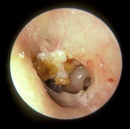
© Michael Hawke MD (Own Work), CC BY 4.0, via Wikimedia Commons
Continuez à lire ci-dessous
Is a cholesteatoma serious?
Retour au sommaireIf left untreated it will expand further and further inside the ear causing permanent hearing loss, through the inner ear and possibly even next to the brain. Where good medical facilities are available, it would be very unusual for it to get that bad but this can happen in regions with limited healthcare facilities.
Types of cholesteatoma
Retour au sommaireCongenital cholesteatoma
This is cholesteatoma which is present at the time of birth. For some reason, even though the eardrum is normal, tiny skin cells get sucked into the middle ear, blocking the Eustachian tube.
This then causes long-term fluid in the middle ear (which should usually be free of fluid) and can cause hearing loss. This becomes apparent between the ages of 6 months to 5 years when the child's hearing does not develop properly. This is a very rare condition and the cause is not fully understood.
Acquired cholesteatoma
This type of cholesteatoma develops later, usually in adults between 30 and 50 years old. Again, the cause is not fully understood. Sometimes a cholesteatoma in an adult can arise after having a grommet, a tiny tube that is put through the eardrum as a treatment for middle ear problems, as a child.
Continuez à lire ci-dessous
Cholesteatoma symptoms
Retour au sommaireA cholesteatoma grows very gradually, over several months, therefore an early cholesteatoma may have no symptoms.
Other common symptoms include:
Écoulement.
Feeling of fullness in the ear.
Discharge
The first symptom is usually a discharge from one ear. It is usually slightly watery, sometimes with a green or yellow colour. The discharge might be slightly smelly and this often looks like an external ear infection (otite externe) or an infection of the inner ear (otite moyenne) with a perforated eardrum when a doctor examines the ear.
Because it looks just like these common infections, it is usually treated (wrongly) with antibiotic ear drops or pills and although it might get slightly better with these treatments, it never fully clears up. A cholesteatoma usually does not cause pain.
Loss of hearing
After a while, hearing loss can occur in that ear. If the cholesteatoma is left untreated it can spread into the balance organ of the inner ear, causing dizziness or unsteadiness. Ringing in the ear (acouphènes) can also occur.
Autres symptômes
Eventually, in very rare cases, it can spread right next to the brain and cause an infection of the brain tissue or the lining of the brain. This is very unlikely to occur as most people would seek medical help if they develop the symptoms described above and the cholesteatoma typically grows very slowly.
Causes of a cholesteatoma
Retour au sommaireWe all have skin inside our ear canal. It is meant to be there and is a normal part of our ear. But rarely, the skin right next to the eardrum, deep in the ear, can get sucked in to the middle ear gradually, where it should not be, resulting in a cholesteatoma.
No one quite knows why this happens but it is usually related to the eardrum being drawn inwards, deeper than it is meant to be (retracted).
This skin then forms a tiny pearl, or ball, that keeps burrowing its way deeper into the ear over many months. It damages the delicate bones inside the middle ear - which are responsible for hearing. At this point it becomes painful.
How common is a cholesteatoma?
Retour au sommaireThis condition is uncommon. Around 7-13 people per 100,000 of the population will be diagnosed with a cholesteatoma each year, and it is estimated that an average doctor will see around one new case every 4-5 years.
Cholesteatoma risk factors
Retour au sommaireCholesteatoma is more common in men than in women and is usually seen in people who have a history of ear infections. Other risk factors include:
Previous surgery.
Ongoing negative pressure in the middle ear, which may cause the eardrum to become retracted ('pulled in').
Trauma to the ear.
Certain genetic problems such as Syndrome de Turner ou syndrome de Down.
How is a cholesteatoma diagnosed?
Retour au sommaireThe doctor or ear specialist (ENT doctor) may suspect a cholesteatoma based on the typical symptoms. When the ear is examined with a torch (an otoscope), the cholesteatoma may be seen. Often there is a hole (perforation) in the eardrum (the tympanic membrane) too.
Because the symptoms come on slowly and mimic common ear infections, the diagnosis is often delayed.
It can be very difficult for a doctor to see a cholesteatoma because usually it causes a lot of pus in the ear which blocks the view to the eardrum.
For this reason the diagnosis is usually made by an ear specialist at a hospital.
The ear specialist will use a tiny suction tube to suck away the discharge and look at the eardrum in detail with a microscope that magnifies the view.
By looking in detail at the eardrum, a specialist can usually see the cholesteatoma pushing through the eardrum.
To then see how far it has spread inside the ear, a specialist scan is needed: this is usually a CT scan or an MRI scan.
Ai-je besoin d'autres tests ?
Retour au sommaireHearing tests (audiometry) may show deafness or hearing loss. These tests are usually performed in a hospital clinic. Samples (swabs) of the ear discharge may also be taken. The discharge often contains a germ (bacterium) called Pseudomonas which is responsible for the smell.
Des Scan CT might be needed to see the extent of the damage caused by the cholesteatoma, and to plan further treatment.
Cholesteatoma treatment
Retour au sommaireAntibiotic eardrops
Antibiotic ear drops can clear away any infection around the cholesteatoma but will not treat the actual problem. Many people will have had antibiotic ear drops prescribed to them without success, before they are diagnosed with a cholesteatoma.
Chirurgie
Surgical treatment is carried out by an ear, nose, and throat specialist (an ENT doctor) and this usually consists of an operation under a general anaesthetic. The aim of the surgery is to remove the cholesteatoma and then clear out part of the middle ear so air can circulate around better. This will hopefully stop the cholesteatoma coming back.
There are different types of cholesteatoma surgery that can be done and a specialist ear doctor will advise which operation is best depending on the size of the cholesteatoma and the patient's medical history.
The commonly performed procedures are a mastoidectomy to clear the cholesteatoma from the bone at the back of the ear, the mastoid, or a 'combined approach tympanoplasty' where the damaged part of the eardrum is also removed and replaced..
Autres traitements
If the patient is not fit for surgery (for example, if they are very old or frail or have other serious medical conditions) then regular visits to an ear specialist will be recommended to suction out any tiny bits of wax or debris deep in the ear. This will not solve the problem but can keep it from getting worse.
Complications of a cholesteatoma
Retour au sommaireUntreated, a cholesteatoma will slowly grow and expand. As it grows it can eat into (erode) and destroy anything in its path.
Possible complications include:
Damage and eventual destruction of the tiny bones of the ear (the ossicles). If these are damaged, permanent deafness can occur.
Damage to the mastoid bone. This is the thick bony lump you can feel behind the ear. The mastoid bone is normally filled with pockets of air (a bit like a honeycomb). A cholesteatoma can grow into the mastoid bone, causing infection and destroying it.
Damage to the cochlea and other structures in the inner ear. This can cause permanent deafness on that side, and/or dizziness and balance problems.
Damage to nearby nerves travelling to the face. This can cause weakness (paralysis) of some of the facial muscles.
Cholesteatoma is often infected and this infection can spread to nearby body parts. Very rarely, a cholesteatoma can erode through the skull next to the ear and into the brain. As a result of spreading infection, conditions such as meningitis and brain abscess may develop. These conditions can cause death.
Please note: although a cholesteatoma sounds nasty, it is not cancerous (malignant) and does not spread to distant parts of the body.
Quelle est la perspective ?
Retour au sommaireThis depends on how much damage has been caused by the cholesteatoma by the time it is found and treated. It is also affected by whether any complications such as meningitis or deafness have occurred. The earlier surgery is done, the better the chance of a good outcome.
If you have had a cholesteatoma, you will need to be followed up in an ENT clinic.
Can a cholesteatoma come back?
If the ear starts discharging again, further surgery may be required. MRI scans are increasingly being used, rather than surgery to review and see whether a cholesteatoma has formed again.
A cholesteatoma grows very gradually, over several months.
Comment entendons-nous ?
Retour au sommaireThe ear is divided into three parts - the external ear, the middle ear and the inner ear. The middle ear, which is behind the eardrum (the tympanic membrane) is filled with air. Air comes from the back of the nose up a thin channel called the Eustachian tube.
In the middle ear there are three tiny bones (ossicles) - the hammer (malleus), anvil (incus) and stirrup (stapes). The inner ear includes the cochlea and the balance organ which contains the semicircular canals, the utricle and saccule.
Sound waves come into the external ear and hit the eardrum. The sound waves cause the eardrum to vibrate. The sound vibrations pass from the eardrum to the ossicles. The ossicles then transmit the vibrations to the cochlea in the inner ear.
The cochlea converts the vibrations to sound signals which are sent along a nerve from the ear to the brain, allowing us to hear.
The semicircular canals and two additional structures called the utricle and saccule in the inner ear all contain a fluid that moves around as we move into different positions. The movement of the fluid is sensed by tiny hairs in the semicircular canals and the utricle and saccule which send messages to the brain along the ear nerve to help maintain balance and posture.
Detail of middle ear
Patient picks for Problèmes d'audition

Oreille, nez et gorge
Maladie de Ménière
La maladie de Ménière provoque généralement des crises de vertiges avec une sensation de rotation (vertige), une perte auditive et des bruits dans l'oreille (acouphènes). Les crises peuvent varier en gravité et en fréquence. Dans certains cas, vous pouvez développer une perte auditive permanente et/ou des acouphènes permanents. Il n'existe pas de traitement curatif, mais une prise en charge peut aider à soulager et à prévenir les symptômes. Si vous êtes conducteur, vous devez arrêter de conduire si un diagnostic de maladie de Ménière est posé. La Direction de l'aviation civile (DVLA) autorisera à nouveau la conduite si les symptômes sont bien contrôlés.
par Dr Doug McKechnie, MRCGP

Oreille, nez et gorge
Cérumen
Une accumulation de cérumen peut entraîner une diminution de l'audition et parfois d'autres symptômes. Le cérumen peut généralement être facilement retiré.
par Dr Philippa Vincent, MRCGP
Lectures complémentaires et références
- Cholestéatome; NICE CKS, août 2024 (accès réservé au Royaume-Uni)
- Castle JT; Cholesteatoma Pearls: Practical Points and Update. Head Neck Pathol. 2018 Sep;12(3):419-429. doi: 10.1007/s12105-018-0915-5. Epub 2018 Aug 1.
- Ayache D, Darrouzet V, Dubrulle F, et al; Imaging of non-operated cholesteatoma: clinical practice guidelines. Eur Ann Otorhinolaryngol Head Neck Dis. 2012 Jun;129(3):148-52. doi: 10.1016/j.anorl.2011.09.005. Epub 2012 Feb 7.
Continuez à lire ci-dessous
About the authorView full bio

Dr Doug McKechnie, MRCGP
Medical Writer
MA, MBBS, MSc, DRCOG, MRCP(UK), MRCGP(2021), FHEA
Dr Doug McKechnie is an NHS GP working in London. He works full-time clinically and is also the Deputy Lead for the Clinical and Professional Practice module at University College London Medical School.
About the reviewerView full bio

Dr Colin Tidy, MRCGP
Médecin généraliste, Auteur médical
MBBS, MRCGP, MRCP (Paediatrics), DCH
Dr Colin Tidy is an NHS Doctor, based in Oxfordshire.
Historique de l'article
Les informations sur cette page sont rédigées et examinées par des cliniciens qualifiés.
Prochaine révision prévue : 2 févr. 2028
3 févr. 2025 | Dernière version

Demandez, partagez, connectez-vous.
Parcourez les discussions, posez des questions et partagez vos expériences sur des centaines de sujets de santé.

Vous ne vous sentez pas bien ?
Évaluez vos symptômes en ligne gratuitement
Inscrivez-vous à la newsletter Patient
Votre dose hebdomadaire de conseils de santé clairs et fiables - rédigés pour vous aider à vous sentir informé, confiant et maître de la situation.
By subscribing you accept our Politique de confidentialité. Vous pouvez vous désabonner à tout moment. Nous ne vendons jamais vos données.