Sténose mitrale
Revu par Dr Adrian Bonsall, MBBSDernière mise à jour par Dr Colin Tidy, MRCGPDernière mise à jour 1 Aug 2017
Respecte les directives éditoriales
- TéléchargerTélécharger
- Partager
- Language
- Discussion
- Version audio
- Ajouter aux sources préférées sur Google
Dans cette série :Maladie des valves cardiaquesRégurgitation mitraleSténose aortiqueRégurgitation aortiqueEndocardite infectieuse
Cette page a été archivée.
Il n'a pas été révisé récemment et n'est pas à jour. Les liens externes et les références peuvent ne plus fonctionner.
La sténose mitrale signifie que lorsque la valve mitrale s'ouvre, elle ne s'ouvre pas complètement. L'ouverture est donc plus étroite que la normale (sténosée).
En un coup d'œil
La sténose mitrale est une condition où la valve mitrale du cœur ne s'ouvre pas complètement.
Cela limite le flux sanguin de l'oreillette gauche au ventricule gauche.
Symptoms can include shortness of breath, tiredness, swollen ankles, and chest pain.
Mild cases may not need treatment, but medication can help ease symptoms.
More severe cases may require surgery to widen or replace the valve.
Note de l'éditeur
Dr Sarah Jarvis, 27th November 2021
NICE guidance on heart valve disease
This leaflet has been archived and has not been reviewed since 2017. It does not include the latest guidance on investigations and treatments for mitral stenosis, which were produced by the National Institute for Health and Care Excellence (NICE) in November 2021.
You can find out more about NICE's recommendations from the further reading section at the end of this leaflet.
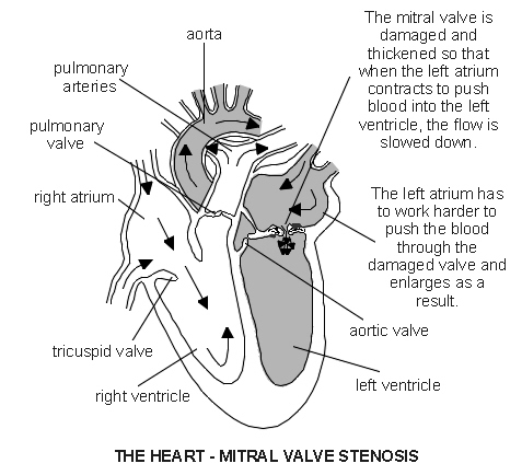
The mitral valve is a heart valve that lies between the left atrium and left ventricle. The valve has two flaps (cusps). The valve allows blood to flow into the left ventricle when the left atrium squeezes (contracts). When the left ventricle contracts, the valve closes and the blood flows out through the aortic valve into the aorta. (The aorta is the main artery which takes blood to the body.)
The cusps are stopped from turning inside out by thin strands of tissue called chordae. The chordae anchor the cusps to the inside wall of the ventricle. The valve or chordae may get damaged or scarred which can prevent the valve from working properly. This can lead to disorders called mitral stenosis, régurgitation mitrale, or both.
Mitral stenosis means that when the mitral valve opens, it does not open fully. The opening is therefore narrower than normal (stenosed). So, there is some restriction of blood flow from the left atrium to the left ventricle. This in turn means there is a reduced amount of blood that is pumped out into the body from the left ventricle. In general, the more narrowed the valve, the less blood can get through and the more severe the problem is likely to be.
Symptômes
If the valve is only mildly narrowed (stenosed) you may have no symptoms or problems. If the stenosis is more severe, the symptoms may include:
Essoufflement, especially with activity or when you lie down
Swollen ankles (œdème)
The heart - mitral valve stenosis
Traitement
Médication
Mild cases may not require any regular medication. Although medicines cannot correct a narrowed (stenosed) mitral valve, some medicines may be prescribed to help ease symptoms, or to help prevent complications - for example, Inhibiteurs de l'enzyme de conversion de l'angiotensine (ECA), 'water tablets' (diuretics) et anticoagulation medication. If you develop fibrillation auriculaire, several medicines can be used to slow the heart rate down.
Traitement chirurgical
Surgical treatment is needed in more severe cases. There are various options, depending of the exact site and severity of the stenosis.
Stretching the stenosed valve
This is a procedure that does not involve open heart surgery. It is called percutaneous balloon commissurotomy or balloon valvuloplasty. (It is called a commissurotomy, as the area where the valve flaps (cusps) come into contact with each other are known as the commissures.)
It is done by inserting a thin tube called a catheter through the skin (percutaneous) into the main blood vessel in the top of the leg. The catheter is passed up to the heart. The tip of the catheter is placed in the mitral valve opening. A balloon at the tip of the catheter is then inflated to stretch the narrowed valve. This is often successful in widening the narrowed valve.
Valve repair is possible in some cases
This is called mitral commissurotomy or mitral valvotomy. This is usually done by open heart surgery. Basically, the edges (commissures) of valve cusps that have become scarred and fused are shaved back to widen the narrowed valve opening.
Valve replacement is needed in some cases
This may be with a mechanical or a tissue valve.
Mechanical valves are made of materials which are not likely to react with your body (for example, those made from titanium), although they can produce a noise which can be heard outside the body.
Tissue valves are made from treated animal tissue (for example, valves from a pig).
If you need surgery, a surgeon will advise on which is the best option for your situation.
What is the outlook for people with mitral stenosis?
In some cases, the disorder is mild and causes no symptoms. If you develop symptoms they tend to become gradually worse over the years. However, the speed of decline can vary. It often takes years for symptoms to become serious. Medication can ease symptoms but cannot reverse a narrowed (stenosed) valve.
Surgical treatments have greatly improved the outlook (prognosis) for most people with more severe stenosis. Surgery has a very good success rate. However, as with all surgical procedures and operations, there is some risk involved when you have surgery. Complications due to surgery occur in a small number of cases.
Sélections des patients pour Maladies cardiaques

Santé cardiaque et vaisseaux sanguins
Régurgitation aortique
L'insuffisance aortique est parfois appelée incompetence aortique ou valve aortique défectueuse. Dans l'insuffisance aortique, la valve ne se ferme pas correctement. La valve aortique est une valve cardiaque située entre le ventricule gauche et l'aorte. Par conséquent, le sang reflue (reflux) dans le ventricule gauche depuis l'aorte. Dans certains cas, l'insuffisance aortique survient en même temps que la sténose aortique. En savoir plus sur la sténose aortique.
par Dr Colin Tidy, MRCGP

Santé cardiaque et vaisseaux sanguins
Syndrome coronarien aigu
Le terme 'syndrome coronarien aigu' (SCA) couvre une gamme de troubles, y compris une crise cardiaque (infarctus du myocarde) et l'angine instable, qui sont causés par une réduction soudaine du flux sanguin vers une partie du muscle cardiaque. Cela est généralement causé par un caillot sanguin.
par Dr Rosalyn Adleman, MRCGP
Questions fréquemment posées
Quelle est la fonction normale de la valve mitrale dans le cœur ?
La valve mitrale est située entre l'oreillette gauche et le ventricule gauche. Son but est de permettre au sang de circuler de l'oreillette gauche vers le ventricule gauche lorsque l'oreillette se contracte. Lorsque le ventricule gauche se contracte ensuite, la valve se ferme, dirigeant le sang à travers la valve aortique vers le reste du corps.
Comment les cordages aident-ils la valve mitrale à fonctionner correctement ?
Les cordages sont de fines bandes de tissu attachées aux volets (cuspides) de la valve mitrale et les ancrent à la paroi interne du ventricule gauche. Leur rôle est crucial pour empêcher les cuspides de la valve de se retourner lorsque le cœur se contracte, assurant ainsi un contrôle adéquat du flux sanguin.
Quelles sont les différences entre les valves mécaniques et les valves tissulaires utilisées pour le remplacement ?
Les valves mécaniques sont fabriquées à partir de matériaux durables comme le titane, qui sont conçus pour ne pas réagir avec le corps, bien qu'elles puissent parfois produire un son audible. Les valves tissulaires, en revanche, sont fabriquées à partir de tissus animaux traités, comme ceux des porcs.
Que se passe-t-il si la valve mitrale ou les cordages sont endommagés ?
Des dommages ou des cicatrices à la valve mitrale ou à ses cordages peuvent empêcher la valve de fonctionner correctement. Cela peut entraîner des conditions telles que la sténose mitrale, où la valve ne s'ouvre pas complètement, ou la régurgitation mitrale, ou les deux, perturbant le flux sanguin normal à travers le cœur.
Lectures complémentaires et références
- Prophylaxie contre l'endocardite infectieuse : Prophylaxie antimicrobienne contre l'endocardite infectieuse chez les adultes et les enfants subissant des procédures interventionnelles; Ligne directrice clinique NICE (mars 2008 - dernière mise à jour juillet 2016)
- 2023 ESC Guidelines for the management of infective endocarditis; European Society of Cardiology (Aug 2023)
- Nishimura RA, Otto CM, Bonow RO, et al; 2017 AHA/ACC Focused Update of the 2014 AHA/ACC Guideline for the Management of Patients With Valvular Heart Disease. Circulation. 2017; CIR.0000000000000503. Originally published March 15, 2017.
- Vahanian A et al; Guidelines on the management of valvular heart disease: The Task Force on the Management of Valvular Heart Disease of the European Society of Cardiology, 2017
- Ozkan M; What is new in ACC/AHA 2017 focused update of valvular heart disease guidelines. Anatol J Cardiol. 2017 Jun;17(6):421-422. doi: 10.14744/AnatolJCardiol.2017.7925.
- Maladie des valves cardiaques chez les adultes : investigation et gestion; Directive NICE (novembre 2021)
À propos de l'auteurVoir la biographie complète

Dr Colin Tidy, MRCGP
Médecin généraliste, Auteur médical
MBBS, MRCGP, MRCP (Paediatrics), DCH
Le Dr Colin Tidy est un médecin du NHS, basé dans l'Oxfordshire.
À propos du critiqueVoir la biographie complète

Dr Adrian Bonsall, MBBS
Auteur Médical
MA (Chimie), MBBS (Hons), DCH
Depuis 2000, Adrian travaille dans les soins pédiatriques d'urgence et de soins intensifs à Sydney, avec des intérêts particuliers pour la toxicologie, le traumatisme et la réanimation.
Historique de l'article
Les informations sur cette page sont rédigées et examinées par des cliniciens qualifiés.
Article également disponible en Anglais, Allemand, Espagnol, Français, Italien, Portugais, Hindi, Hébreu, Arabe, and Suédois.
1 Aug 2017 | Dernière version

Demandez, partagez, connectez-vous.
Parcourez les discussions, posez des questions et partagez vos expériences sur des centaines de sujets de santé.

Vous ne vous sentez pas bien ?
Évaluez vos symptômes en ligne gratuitement
Inscrivez-vous à la newsletter Patient
Votre dose hebdomadaire de conseils de santé clairs et fiables - rédigés pour vous aider à vous sentir informé, confiant et maître de la situation.
En vous abonnant, vous acceptez notre Politique de confidentialité. Vous pouvez vous désabonner à tout moment. Nous ne vendons jamais vos données.