Barium tests
Peer reviewed by Dr Surangi Mendis, MRCGPLast updated by Dr Rosalyn Adleman, MRCGPLast updated 17 Aug 2023
Meets Patient’s editorial guidelines
- DownloadDownload
- Share
- Language
- Discussion
- Audio Version
- Add to preferred sources on Google
Barium tests are used to help see the outline of various parts of the gut (gastrointestinal tract). These include the gullet (oesophagus), stomach, upper gut (small intestines) and colon (large intestine).
Barium X-ray tests are done less commonly these days. Today we usually look into the gut with a flexible telescope (endoscopy or colonoscopy) . However, there is still a place for barium tests to help assess various problems of the gut.
Note: the information below is a general guide only. The arrangements, and the way tests are performed, may vary between different hospitals. Always follow the instructions given by your doctor or local hospital.
At a glance
Barium tests use a special liquid to make parts of your gut show up on X-rays.
Types of barium tests include swallow, meal, follow through, small intestine enema, and enema.
You will usually be asked not to eat for several hours before the test.
After the test, drink fluids and eat fibre to help prevent constipation from the barium.
Barium will make your poo white until it has all left your system.
Tell your doctor if you are pregnant or have insulin-dependent diabetes.
Why is barium used during some X-ray tests?
The gut (gastrointestinal tract) does not show up very well on ordinary X-ray pictures. However, if you drink a white liquid that contains a chemical called barium sulfate, the outline of the upper parts of the gut (oesophagus, stomach and small intestines) shows up clearly on X-ray pictures. Similarly, if barium is placed in the colon this can also be seen clearly on X-ray. This is because X-rays do not pass through barium.
Types of barium test
Diagram naming the parts of the gut
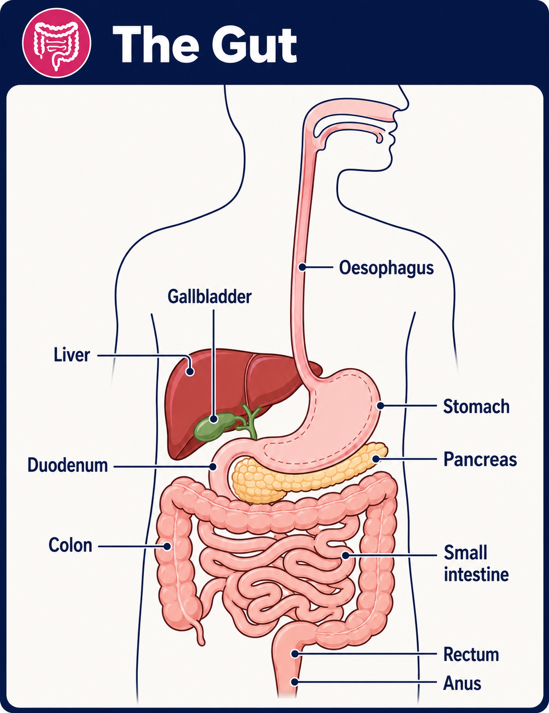
Depending on what part of your gut is being looked at, you may have one or more of the tests listed below. In each test, the barium coats the lining of the gut being tested. Therefore, abnormalities in the lining or structure of the gut can be seen on the X-ray pictures.
In each of the following tests, several X-ray pictures are taken using low-dose X-rays. The total amount of radiation for each test is quite small and thought to be safe. The X-ray machine is usually linked to a TV monitor. Still pictures, or a video recording of X-ray pictures taken in quick succession, can be taken if necessary.
Barium swallow
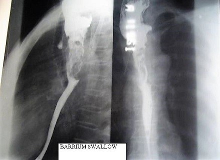
In this test you drink some barium liquid. The barium liquid is often fruit-flavoured so it is pleasant to drink. You stand in front of an X-ray machine whilst X-ray pictures are taken as you swallow. This test aims to look for problems in the gullet.
These include a narrowing (stricture), hiatus hernias, tumours, reflux from the stomach, disorders of swallowing, etc. You will usually be asked not to eat or drink for a few hours before this test. A barium swallow test takes about 10 minutes.
Barium swallow X-ray
Barium meal
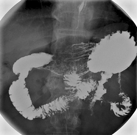
This is similar to a barium swallow. However, it aims to look for problems in the stomach and the first part of the gut (small intestine), known as the duodenum. These problems may include ulcers, small fleshy lumps (polyps), tumours, etc.
You drink some barium liquid but you then lie on a couch whilst X-ray pictures are taken over your tummy. It may take a little longer to do than a barium swallow.
So that the barium coats all around the lining of the stomach, the doctor doing the test (radiologist) may do one or more of the following:
Ask you to swallow some bicarbonate powder and citric acid before swallowing the barium. These 'fizz up' when they mix in the stomach and make some gas. (You may have the urge to burp.) The gas expands the stomach and duodenum and also pushes the barium to coat the lining of the stomach and duodenum. This makes the X-ray pictures clearer. It is the shape and contours of the lining of the stomach and duodenum which need to be seen most clearly on the pictures.
Ask you to turn over on to your stomach on the couch. Various X-ray pictures may be taken whilst you are in different positions.
You may be given an injection of a drug that makes the muscles in the stomach and gut relax.
Can I eat before a barium meal?
You will usually be asked not to eat anything for several hours before this test. (Food particles in the gut can make it difficult to interpret the X-rays). However, you may be allowed sips of water up to two hours before the test.
Barium meal stomach X-ray
Barium follow through
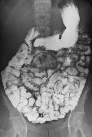
This test is similar to a barium meal but aims to look for problems in the small intestine. Therefore, you drink the barium liquid but then need to wait 10-15 minutes before any X-rays are taken. This allows time for the barium to reach the small intestine.
You may then have an X-ray every 30 minutes or so until the barium is seen to have gone through all the small intestine and reached the large intestine (colon). This test will last longer than the previous ones. The overall time taken will depend on how quickly your gut moves things along.
Barium follow through showing small bowel
Small intestine enema
This test is similar to a barium follow through. However, instead of drinking the barium liquid, a thin tube is passed down your gullet, through the stomach and into the first part of the small intestine.
Barium liquid is then poured down the tube. This test is not commonly done but can give some different information about the small intestine to the tests above.
Barium enema
This is a test to take X-ray pictures of the colon. Instead of drinking the barium it is placed in your colon (large intestine). You will be asked to lie on a couch and a small tube is put into your back passage (anus) and gently pushed up a few centimetres. Barium liquid is then passed through the tube into your colon (large intestine). So that the barium coats all of the lining of the colon, the doctor doing the test may be do some of the following:
Ask you to change position on the couch, for example on your side or back.
Give you an injection of a drug that makes the muscles in the walls of the colon relax.
Pass some air down the enema tube into the colon. This may feel a little uncomfortable, like trapped wind. But it helps makes the x-ray pictures much clearer.
When the barium liquid has spread throughout the colon, several x-ray pictures are taken so that all parts of the colon can be seen. The tube is then removed and you can go to the toilet. This test takes around 15-20 minutes.
What preparation do I need to do before a barium test?
This will depend on which of the tests listed above you need to have. Your local hospital will give you advice on what to do before the procedure.
After you have had a barium X-ray test
You should be able to go home as soon as the test is finished.
You can eat normally straight after any barium test.
The barium does not get absorbed into the body. Therefore, it is rare for a barium test to cause any other complications or side-effects.
Are there any side-effects of a barium x-ray?
Some people feel a little sickly for a few hours afterwards.
The barium may make you constipated. Therefore, to help prevent constipation:
Have lots to drink for a day or so to flush the barium out of your gut.
Eat plenty of fruit and vegetables for a day or so.
See your doctor if you haven't passed any poo after three or four days.
The barium will make your poo white or pale until it has all come out of your gut (after a day or so).
If you had an injection to relax the muscles in your stomach, it may cause some blurring of your vision for an hour or so. If this happens it is best not to drive.
Some other points about barium X-ray tests
Tell your doctor if you have insulin-dependent diabetes, so that you can arrange for the best time for you to stop eating and for the test to be done.
Pregnant women, if possible, should not have an X-ray test, as there is a small risk that X-rays may harm the unborn child. This is why women are asked before having an X-ray whether they are, or might be, pregnant.
Patient picks for Imaging
Tests and investigations
PET scan
A PET scan create images which show where cells are particularly active in the body. It is most commonly used to diagnose and assess cancer. Note: the information below is a general guide only. The arrangements, and the way tests are performed, may vary between different hospitals. Always follow the instructions given by your doctor or local hospital.
by Dr Rosalyn Adleman, MRCGP

Tests and investigations
CT scan
Note: the information below is a general guide only. The arrangements, and the way tests are performed, may vary between different hospitals. Always follow the instructions given by your doctor or local hospital.
by Dr Rachel Hudson, MRCGP
Frequently asked questions
What is barium?
Barium is a chemical that, when used in the form of barium sulfate, helps the gut show up clearly on X-ray pictures. X-rays do not pass through barium, so it coats the lining of the gut, making its outline visible.
Does barium make you poop?
The barium itself is not absorbed into your body. It will pass through your digestive system and make your stool (poo) appear white or pale until it has all left your gut, which typically happens after a day or so.
Does barium sulfate make you sick?
Some people might feel a little sickly for a few hours after a barium test. However, it's rare for barium tests to cause complications or other side-effects because the barium is not absorbed by the body.
How long does barium stay in your system?
Barium typically stays in your gut for about a day or so. You can help flush it out quickly by drinking plenty of fluids and eating lots of fruit and vegetables after the test. If you haven't passed any stool after three or four days, you should consult your doctor.
Can I drive after a barium X-ray test?
You can usually go home as soon as the test is finished. However, if you received an injection to relax the muscles in your stomach, it might cause temporarily blurred vision for about an hour. If this happens, it is best not to drive until your vision returns to normal.
Further reading and references
- Nin CS, Marchiori E, Irion KL, et al; Barium swallow study in routine clinical practice: a prospective study in patients with chronic cough. J Bras Pneumol. 2013 Nov-Dec;39(6):686-91. doi: 10.1590/S1806-37132013000600007.
- Negreanu L, Smarandache G, Mateescu RB; Role of capsule endoscopy Pillcam COLON 2 in patients with known or suspected Crohn's disease who refused colonoscopy or underwent incomplete colonoscopic exam: a case series. Tech Coloproctol. 2014 Mar;18(3):277-83. doi: 10.1007/s10151-013-1054-3. Epub 2013 Aug 21.
- Li YZ, Wu PH; Conventional radiological strategy of common gastrointestinal neoplasms. World J Radiol. 2015 Jan 28;7(1):7-16. doi: 10.4329/wjr.v7.i1.7.
About the authorView full bio
Updated by

Dr Rosalyn Adleman, MRCGP
MRCGP
Dr Rosalyn Adleman, is an NHS GP working in north London.
About the reviewerView full bio

Dr Surangi Mendis, MRCGP
Consultant and Medical Author
MBBS, BSc (1st), MRCGP (2014), DFSRH, PGcert otology and audiology
Surangi Mendis is a consultant in Audiovestibular Medicine and Neuro-otology at The Royal National ENT and Eastman Dental Hospitals, UCLH.
Join the conversation
Share your thoughts or experiences with others in our patient communities.

Ask, share, connect.
Browse discussions, ask questions, and share experiences across hundreds of health topics.

Feeling unwell?
Assess your symptoms online for free
Sign up to the Patient newsletter
Your weekly dose of clear, trustworthy health advice - written to help you feel informed, confident and in control.
By subscribing you accept our Privacy Policy. You can unsubscribe at any time. We never sell your data.